Show PDB file:   
         Plain Text   HTML   (compressed file size)
QuickSearch:   
by PDB,NDB,UniProt,PROSITE Code or Search Term(s)  
(-)Asym./Biol. Unit
(-)Asym./Biol. Unit - sites
collapse expand < >
Image Asym./Biol. Unit
Asym./Biol. Unit  (Jmol Viewer)
Image Asym./Biol. Unit - sites
Asym./Biol. Unit - sites  (Jmol Viewer)

(-) Description

Title :  X-RAY STRUCTURE OF MCPS LIGAND BINDING DOMAIN IN COMPLEX WITH MALATE
 
Authors :  E. Pineda-Molina, J. A. Gavira, T. Krell
Date :  05 Apr 11  (Deposition) - 18 Apr 12  (Release) - 10 Jul 13  (Revision)
Method :  X-RAY DIFFRACTION
Resolution :  1.80
Chains :  Asym./Biol. Unit :  A,B
Keywords :  Receptor, Chemoreceptor, Chemotaxis (Keyword Search: [Gene Ontology, PubMed, Web (Google))
 
Reference :  E. Pineda-Molina, J. Reyes-Darias, J. Lacal, J. L. Ramos, J. M. Garcia-Ruiz, J. A. Gavira, T. Krell
Evidence For Chemoreceptors With Bimodular Ligand-Binding Regions Harboring Two Signal-Binding Sites.
Proc. Natl. Acad. Sci. Usa V. 109 18926 2012
PubMed-ID: 23112148  |  Reference-DOI: 10.1073/PNAS.1201400109
(for further references see the PDB file header)

(-) Compounds

Molecule 1 - METHYL-ACCEPTING CHEMOTAXIS TRANSDUCER
    ChainsA, B
    EngineeredYES
    Expression SystemESCHERICHIA COLI
    Expression System StrainBL21(DE3)
    Expression System Taxid469008
    FragmentLIGAND BINDING DOMAIN, RESIDUES 46-283
    Organism ScientificPSEUDOMONAS PUTIDA
    Organism Taxid160488
    StrainKT2440
    SynonymMCPS

 Structural Features

(-) Chains, Units

  12
Asymmetric/Biological Unit AB

Summary Information (see also Sequences/Alignments below)
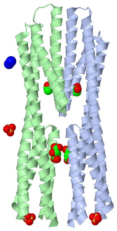
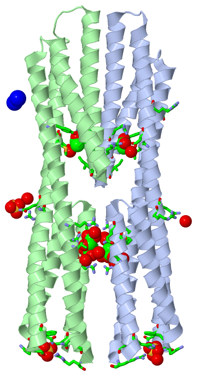
(-) Ligands, Modified Residues, Ions  (4, 9)

Asymmetric/Biological Unit (4, 9)
No.NameCountTypeFull Name
1ACT2Ligand/IonACETATE ION
2LMR2Ligand/Ion(2S)-2-HYDROXYBUTANEDIOIC ACID
3NA2Ligand/IonSODIUM ION
4SO43Ligand/IonSULFATE ION

(-) Sites  (9, 9)

Asymmetric Unit (9, 9)
No.NameEvidenceResiduesDescription
1AC1SOFTWAREARG A:64 , ARG A:67 , ARG A:258 , THR A:262 , HOH A:2254 , HOH A:2272 , HOH A:2273 , HOH A:2274 , HOH A:2275 , GLN B:69BINDING SITE FOR RESIDUE LMR A 501
2AC2SOFTWAREMET A:141 , ASP A:142 , ARG A:187 , VAL A:190 , ARG A:191 , ILE A:194 , TYR A:240 , HOH A:2167BINDING SITE FOR RESIDUE ACT A 503
3AC3SOFTWAREGLN A:69 , ARG B:64 , ARG B:67 , ARG B:258 , THR B:262 , HOH B:2254 , HOH B:2255 , HOH B:2256 , HOH B:2257BINDING SITE FOR RESIDUE LMR B 501
4AC4SOFTWAREMET B:141 , ASP B:142 , ARG B:187 , VAL B:190 , ARG B:191 , ILE B:194 , TYR B:240 , HOH B:2153BINDING SITE FOR RESIDUE ACT B 504
5AC5SOFTWAREHIS A:49 , PHE A:101 , LYS A:102 , SER A:103 , ASN A:106 , GLN A:279 , ARG A:282 , HOH A:2276BINDING SITE FOR RESIDUE SO4 A1283
6AC6SOFTWAREHIS B:49 , PHE B:101 , LYS B:102 , SER B:103 , ASN B:106 , GLN B:279 , ARG B:282BINDING SITE FOR RESIDUE SO4 A1284
7AC7SOFTWAREARG A:128 , GLN A:129 , HOH A:2133 , ARG B:128 , GLN B:129 , HOH B:2129BINDING SITE FOR RESIDUE SO4 B1283
8AC8SOFTWAREARG A:147BINDING SITE FOR RESIDUE NA B1284
9AC9SOFTWAREASN A:235BINDING SITE FOR RESIDUE NA B1285

(-) SS Bonds  (0, 0)

(no "SS Bond" information available for 2YFA)

(-) Cis Peptide Bonds  (0, 0)

(no "Cis Peptide Bond" information available for 2YFA)

 Sequence-Structure Mapping

(-) SAPs(SNPs)/Variants  (0, 0)

(no "SAP(SNP)/Variant" information available for 2YFA)

(-) PROSITE Motifs  (0, 0)

(no "PROSITE Motif" information available for 2YFA)

(-) Exons   (0, 0)

(no "Exon" information available for 2YFA)

(-) Sequences/Alignments

Asymmetric/Biological Unit
   Reformat: Number of residues per line =  ('0' or empty: single-line sequence representation)
  Number of residues per labelling interval =   
  UniProt sequence: complete  aligned part    
   Show mapping: SCOP domains CATH domains Pfam domains Secondary structure (by author)
SAPs(SNPs) PROSITE motifs Exons
(details for a mapped element are shown in a popup box when the mouse pointer rests over it)
Chain A from PDB  Type:PROTEIN  Length:236
 aligned with MCPS_PSEPK | Q88E10 from UniProtKB/Swiss-Prot  Length:639

    Alignment length:236
                                    52        62        72        82        92       102       112       122       132       142       152       162       172       182       192       202       212       222       232       242       252       262       272      
           MCPS_PSEPK    43 SNWMGDIGQLNKDLTDLRIARLQYMIANGDDTAAANTLAKLDAFSKQQAYLATTFKSPENVKLLGELGDTISAYKLSLNKMRQGYDATRAARVSMDSSAIRADQAMDALSQEVMARPEADSVRLAQYQLISKARQQLLQVRIDVRGYIAENSSANEQAALRQLDAALADTDNLKRQLPSEDARLQQFENAVLAYRDAVRQFRDAVANITTSRAEMTVQGADIVKRSDALYQIQLER 278
               SCOP domains -------------------------------------------------------------------------------------------------------------------------------------------------------------------------------------------------------------------------------------------- SCOP domains
               CATH domains -------------------------------------------------------------------------------------------------------------------------------------------------------------------------------------------------------------------------------------------- CATH domains
               Pfam domains -------------------------------------------------------------------------------------------------------------------------------------------------------------------------------------------------------------------------------------------- Pfam domains
         Sec.struct. author hhhhhhhhhhhhhhhhhhhhhhhhhhh..hhhhhhhhhhhhhhhhhhhhhhhhhh.hhhhhhhhhhhhhhhhhhhhhhhhhhhhhhhhhhhhhhhhhhhhhhhhhhhhhhhhhhhh..hhhhhhhhhhhhhhhhhhhhhhhhhhhhhhhh.hhhhhhhhhhhhhhhhhhhhhhhhhh..hhhhhhhhhhhhhhhhhhhhhhhhhhhhhhhhhhhhhhhhhhhhhhhhhhhhhhhh. Sec.struct. author
                 SAPs(SNPs) -------------------------------------------------------------------------------------------------------------------------------------------------------------------------------------------------------------------------------------------- SAPs(SNPs)
                    PROSITE -------------------------------------------------------------------------------------------------------------------------------------------------------------------------------------------------------------------------------------------- PROSITE
                 Transcript -------------------------------------------------------------------------------------------------------------------------------------------------------------------------------------------------------------------------------------------- Transcript
                 2yfa A  47 GSHMGDIGQLNKDLTDLRIARLQYMIANGDDTAAANTLAKLDAFSKQQAYLATTFKSPENVKLLGELGDTISAYKLSLNKMRQGYDATRAARVSMDSSAIRADQAMDALSQEVMARPEADSVRLAQYQLISKARQQLLQVRIDVRGYIAENSSANEQAALRQLDAALADTDNLKRQLPSEDARLQQFENAVLAYRDAVRQFRDAVANITTSRAEMTVQGADIVKRSDALYQIQLER 282
                                    56        66        76        86        96       106       116       126       136       146       156       166       176       186       196       206       216       226       236       246       256       266       276      

Chain B from PDB  Type:PROTEIN  Length:235
 aligned with MCPS_PSEPK | Q88E10 from UniProtKB/Swiss-Prot  Length:639

    Alignment length:235
                                    53        63        73        83        93       103       113       123       133       143       153       163       173       183       193       203       213       223       233       243       253       263       273     
           MCPS_PSEPK    44 NWMGDIGQLNKDLTDLRIARLQYMIANGDDTAAANTLAKLDAFSKQQAYLATTFKSPENVKLLGELGDTISAYKLSLNKMRQGYDATRAARVSMDSSAIRADQAMDALSQEVMARPEADSVRLAQYQLISKARQQLLQVRIDVRGYIAENSSANEQAALRQLDAALADTDNLKRQLPSEDARLQQFENAVLAYRDAVRQFRDAVANITTSRAEMTVQGADIVKRSDALYQIQLER 278
               SCOP domains ------------------------------------------------------------------------------------------------------------------------------------------------------------------------------------------------------------------------------------------- SCOP domains
               CATH domains ------------------------------------------------------------------------------------------------------------------------------------------------------------------------------------------------------------------------------------------- CATH domains
               Pfam domains ------------------------------------------------------------------------------------------------------------------------------------------------------------------------------------------------------------------------------------------- Pfam domains
         Sec.struct. author hhhhhhhhhhhhhhhhhhhhhhhhhh..hhhhhhhhhhhhhhhhhhhhhhhhhh.hhhhhhhhhhhhhhhhhhhhhhhhhhhhhhhhhhhhhhhhhhhhhhhhhhhhhhhh......hhhhhhhhhhhhhhhhhhhhhhhhhhhhhhhh.hhhhhhhhhhhhhhhhhhhhhhhhhh..hhhhhhhhhhhhhhhhhhhhhhhhhhhhhhhhhhhhhhhhhhhhhhhhhhhhhhhh. Sec.struct. author
                 SAPs(SNPs) ------------------------------------------------------------------------------------------------------------------------------------------------------------------------------------------------------------------------------------------- SAPs(SNPs)
                    PROSITE ------------------------------------------------------------------------------------------------------------------------------------------------------------------------------------------------------------------------------------------- PROSITE
                 Transcript ------------------------------------------------------------------------------------------------------------------------------------------------------------------------------------------------------------------------------------------- Transcript
                 2yfa B  48 SHMGDIGQLNKDLTDLRIARLQYMIANGDDTAAANTLAKLDAFSKQQAYLATTFKSPENVKLLGELGDTISAYKLSLNKMRQGYDATRAARVSMDSSAIRADQAMDALSQEVMARPEADSVRLAQYQLISKARQQLLQVRIDVRGYIAENSSANEQAALRQLDAALADTDNLKRQLPSEDARLQQFENAVLAYRDAVRQFRDAVANITTSRAEMTVQGADIVKRSDALYQIQLER 282
                                    57        67        77        87        97       107       117       127       137       147       157       167       177       187       197       207       217       227       237       247       257       267       277     

   Legend:   → Mismatch (orange background)
  - → Gap (green background, '-', border residues have a numbering label)
    → Modified Residue (blue background, lower-case, 'x' indicates undefined single-letter code, labelled with number + name)
  x → Chemical Group (purple background, 'x', labelled with number + name, e.g. ACE or NH2)
  extra numbering lines below/above indicate numbering irregularities and modified residue names etc., number ends below/above '|'

 Classification and Annotation

(-) SCOP Domains  (0, 0)

(no "SCOP Domain" information available for 2YFA)

(-) CATH Domains  (0, 0)

(no "CATH Domain" information available for 2YFA)

(-) Pfam Domains  (0, 0)

(no "Pfam Domain" information available for 2YFA)

(-) Gene Ontology  (6, 6)

Asymmetric/Biological Unit(hide GO term definitions)
Chain A,B   (MCPS_PSEPK | Q88E10)
molecular function
    GO:0004871    signal transducer activity    Conveys a signal across a cell to trigger a change in cell function or state. A signal is a physical entity or change in state that is used to transfer information in order to trigger a response.
biological process
    GO:0006935    chemotaxis    The directed movement of a motile cell or organism, or the directed growth of a cell guided by a specific chemical concentration gradient. Movement may be towards a higher concentration (positive chemotaxis) or towards a lower concentration (negative chemotaxis).
    GO:0007165    signal transduction    The cellular process in which a signal is conveyed to trigger a change in the activity or state of a cell. Signal transduction begins with reception of a signal (e.g. a ligand binding to a receptor or receptor activation by a stimulus such as light), or for signal transduction in the absence of ligand, signal-withdrawal or the activity of a constitutively active receptor. Signal transduction ends with regulation of a downstream cellular process, e.g. regulation of transcription or regulation of a metabolic process. Signal transduction covers signaling from receptors located on the surface of the cell and signaling via molecules located within the cell. For signaling between cells, signal transduction is restricted to events at and within the receiving cell.
cellular component
    GO:0016021    integral component of membrane    The component of a membrane consisting of the gene products and protein complexes having at least some part of their peptide sequence embedded in the hydrophobic region of the membrane.
    GO:0016020    membrane    A lipid bilayer along with all the proteins and protein complexes embedded in it an attached to it.
    GO:0005886    plasma membrane    The membrane surrounding a cell that separates the cell from its external environment. It consists of a phospholipid bilayer and associated proteins.

 Visualization

(-) Interactive Views

Asymmetric/Biological Unit
  Complete Structure
    Jena3D(integrated viewing of ligand, site, SAP, PROSITE, SCOP information)
    WebMol | AstexViewer[tm]@PDBe
(Java Applets, require no local installation except for Java; loading may be slow)
    STRAP
(Java WebStart application, automatic local installation, requires Java; full application with system access!)
    RasMol
(require local installation)
    Molscript (VRML)
(requires installation of a VRML viewer; select preferred view via VRML and generate a mono or stereo PDF format file)
 
  Ligands, Modified Residues, Ions
    ACT  [ RasMol | Jena3D ]  +environment [ RasMol | Jena3D ]
    LMR  [ RasMol | Jena3D ]  +environment [ RasMol | Jena3D ]
    NA  [ RasMol | Jena3D ]  +environment [ RasMol | Jena3D ]
    SO4  [ RasMol | Jena3D ]  +environment [ RasMol | Jena3D ]
 
  Sites
    AC1  [ RasMol ]  +environment [ RasMol ]
    AC2  [ RasMol ]  +environment [ RasMol ]
    AC3  [ RasMol ]  +environment [ RasMol ]
    AC4  [ RasMol ]  +environment [ RasMol ]
    AC5  [ RasMol ]  +environment [ RasMol ]
    AC6  [ RasMol ]  +environment [ RasMol ]
    AC7  [ RasMol ]  +environment [ RasMol ]
    AC8  [ RasMol ]  +environment [ RasMol ]
    AC9  [ RasMol ]  +environment [ RasMol ]
 
  Cis Peptide Bonds
(no "Cis Peptide Bonds" information available for 2yfa)
 

(-) Still Images

Jmol
  protein: cartoon or spacefill or dots and stick; nucleic acid: cartoon and stick; ligands: spacefill; active site: stick
Molscript
  protein, nucleic acid: cartoon; ligands: spacefill; active site: ball and stick

 Databases and Analysis Tools

(-) Databases

Access by PDB/NDB ID
  2yfa
    Family and Domain InformationProDom | SYSTERS
    General Structural InformationGlycoscienceDB | MMDB | NDB | OCA | PDB | PDBe | PDBj | PDBsum | PDBWiki | PQS | PROTEOPEDIA
    Orientation in MembranesOPM
    Protein SurfaceSURFACE
    Secondary StructureDSSP (structure derived) | HSSP (homology derived)
    Structural GenomicsGeneCensus
    Structural NeighboursCE | VAST
    Structure ClassificationCATH | Dali | SCOP
    Validation and Original DataBMRB Data View | BMRB Restraints Grid | EDS | PROCHECK | RECOORD | WHAT_CHECK
 
Access by UniProt ID/Accession number
  MCPS_PSEPK | Q88E10
    Comparative Protein Structure ModelsModBase
    Genomic InformationEnsembl
    Protein-protein InteractionDIP
    Sequence, Family and Domain InformationInterPro | Pfam | SMART | UniProtKB/SwissProt
 
Access by Enzyme Classificator   (EC Number)
  (no 'Enzyme Classificator' available)
    General Enzyme InformationBRENDA | EC-PDB | Enzyme | IntEnz
    PathwayKEGG | MetaCyc
 
Access by Disease Identifier   (MIM ID)
  (no 'MIM ID' available)
    Disease InformationOMIM
 
Access by GenAge ID
  (no 'GenAge ID' available)
    Age Related InformationGenAge

(-) Analysis Tools

Access by PDB/NDB ID
    Domain InformationXDom
    Interatomic Contacts of Structural UnitsCSU
    Ligand-protein ContactsLPC
    Protein CavitiescastP
    Sequence and Secondary StructurePDBCartoon
    Structure AlignmentSTRAP(Java WebStart application, automatic local installation, requires Java; full application with system access!)
    Structure and Sequence BrowserSTING
 
Access by UniProt ID/Accession number
  MCPS_PSEPK | Q88E10
    Protein Disorder PredictionDisEMBL | FoldIndex | GLOBPLOT (for more information see DisProt)

 Related Entries

(-) Entries Sharing at Least One Protein Chain (UniProt ID)

UniProtKB/Swiss-Prot
        MCPS_PSEPK | Q88E102yfb

(-) Related Entries Specified in the PDB File

2yfb X-RAY STRUCTURE OF MCPS LIGAND BINDING DOMAIN IN COMPLEX WITH SUCCINATE