Show PDB file:   
         Plain Text   HTML   (compressed file size)
QuickSearch:   
by PDB,NDB,UniProt,PROSITE Code or Search Term(s)  
(-)Asymmetric Unit
(-)Asym. Unit - sites
(-)Biological Unit 1
(-)Biol. Unit 1 - sites
collapse expand < >
Image Asymmetric Unit
Asymmetric Unit  (Jmol Viewer)
Image Asym. Unit - sites
Asym. Unit - sites  (Jmol Viewer)
Image Biological Unit 1
Biological Unit 1  (Jmol Viewer)
Image Biol. Unit 1 - sites
Biol. Unit 1 - sites  (Jmol Viewer)

(-) Description

Title :  THE STRUCTURE OF A D-LYXOSE ISOMERASE FROM THE SIGMAB REGULON OF BACILLUS SUBTILIS
 
Authors :  J. Marles-Wright, R. J. Lewis
Date :  07 Dec 10  (Deposition) - 25 May 11  (Release) - 25 May 11  (Revision)
Method :  X-RAY DIFFRACTION
Resolution :  1.23
Chains :  Asym. Unit :  A
Biol. Unit 1:  A  (2x)
Keywords :  Isomerase, Carbohydrate Metabolism, Metal-Binding, Sugar Isomerase, Stress Response (Keyword Search: [Gene Ontology, PubMed, Web (Google))
 
Reference :  J. Marles-Wright, R. J. Lewis
The Structure Of A D-Lyxose Isomerase From The Sigmab Regulon Of Bacillus Subtilis
Proteins V. 79 2015 2011
PubMed-ID: 21520290  |  Reference-DOI: 10.1002/PROT.23028

(-) Compounds

Molecule 1 - PROBABLE D-LYXOSE KETOL-ISOMERASE
    ChainsA
    EC Number5.3.1.15
    EngineeredYES
    Expression SystemESCHERICHIA COLI
    Expression System PlasmidPET28B
    Expression System StrainB834
    Expression System Taxid562
    Expression System Vector TypePLASMID
    Organism ScientificBACILLUS SUBTILIS SUBSP. SUBTILIS
    Organism Taxid224308
    Other DetailsZINC AND ARSENIC COORDINATION IN ACTIVE SITE
    Strain168

 Structural Features

(-) Chains, Units

  1
Asymmetric Unit A
Biological Unit 1 (2x)A

Summary Information (see also Sequences/Alignments below)
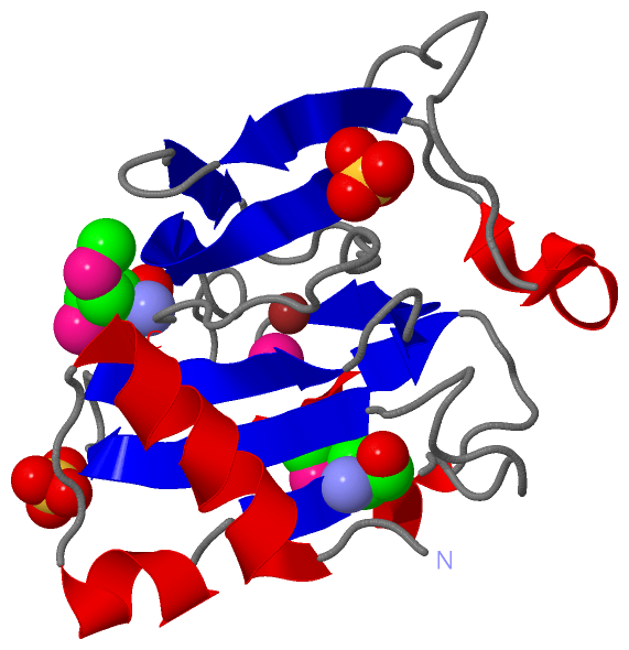
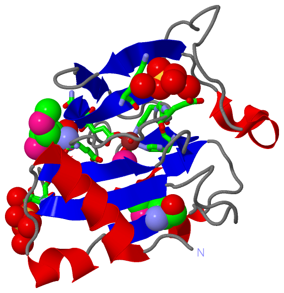
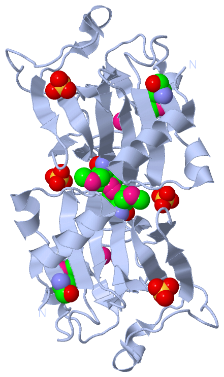
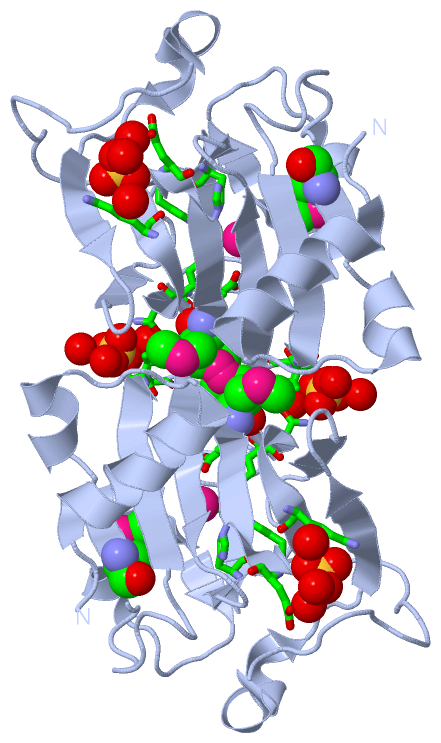
(-) Ligands, Modified Residues, Ions  (4, 6)

Asymmetric Unit (4, 6)
No.NameCountTypeFull Name
1ARS1Ligand/IonARSENIC
2MSE2Mod. Amino AcidSELENOMETHIONINE
3SO42Ligand/IonSULFATE ION
4ZN1Ligand/IonZINC ION
Biological Unit 1 (3, 10)
No.NameCountTypeFull Name
1ARS2Ligand/IonARSENIC
2MSE4Mod. Amino AcidSELENOMETHIONINE
3SO44Ligand/IonSULFATE ION
4ZN-1Ligand/IonZINC ION

(-) Sites  (4, 4)

Asymmetric Unit (4, 4)
No.NameEvidenceResiduesDescription
1AC1SOFTWAREHIS A:69 , HIS A:71 , GLU A:82 , HIS A:137 , ARS A:1176 , HOH A:2262 , HOH A:2263BINDING SITE FOR RESIDUE ZN A1173
2AC2SOFTWARELYS A:90 , TYR A:92 , GLY A:142 , GLU A:143 , HOH A:2264 , HOH A:2265 , HOH A:2266BINDING SITE FOR RESIDUE SO4 A1174
3AC3SOFTWARETHR A:50 , ASP A:51 , GLN A:81 , HOH A:2116 , HOH A:2267 , HOH A:2268BINDING SITE FOR RESIDUE SO4 A1175
4AC4SOFTWAREGLU A:150 , ZN A:1173 , HOH A:2234 , HOH A:2263BINDING SITE FOR RESIDUE ARS A1176

(-) SS Bonds  (0, 0)

(no "SS Bond" information available for 2Y0O)

(-) Cis Peptide Bonds  (0, 0)

(no "Cis Peptide Bond" information available for 2Y0O)

 Sequence-Structure Mapping

(-) SAPs(SNPs)/Variants  (0, 0)

(no "SAP(SNP)/Variant" information available for 2Y0O)

(-) PROSITE Motifs  (0, 0)

(no "PROSITE Motif" information available for 2Y0O)

(-) Exons   (0, 0)

(no "Exon" information available for 2Y0O)

(-) Sequences/Alignments

Asymmetric Unit
   Reformat: Number of residues per line =  ('0' or empty: single-line sequence representation)
  Number of residues per labelling interval =   
  UniProt sequence: complete  aligned part    
   Show mapping: SCOP domains CATH domains Pfam domains Secondary structure (by author)
SAPs(SNPs) PROSITE motifs Exons
(details for a mapped element are shown in a popup box when the mouse pointer rests over it)
Chain A from PDB  Type:PROTEIN  Length:171
 aligned with DLYKI_BACSU | P96578 from UniProtKB/Swiss-Prot  Length:167

    Alignment length:171
                                                                                                                                                                                               167     
                                    11        21        31        41        51        61        71        81        91       101       111       121       131       141       151       161     |   - 
          DLYKI_BACSU     2 GITKEEVNSYYQKAGIVLTDEEVDQIQLMDYGLGKERKVGLQLFVYVNTDRYCSKELVLFPGQTCPEHRHPPVDGQEGKQETFRCRYGKVYLYVEGEKTPLPKVLPPQEDREHYTVWHEIELEPGGQYTIPPNTKHWFQAGEEGAVVTEMSSTSTDKHDIFTDPRI-----   -
               SCOP domains --------------------------------------------------------------------------------------------------------------------------------------------------------------------------- SCOP domains
               CATH domains --------------------------------------------------------------------------------------------------------------------------------------------------------------------------- CATH domains
               Pfam domains --------------------------------------------------------------------------------------------------------------------------------------------------------------------------- Pfam domains
         Sec.struct. author ..hhhhhhhhhhhh....hhhhhh..eee...........eeeeeeee...eeeeeeee....eeeeee...........eeeeeeeeeeeeee............hhhhhhhh...eeeee....eeee.....eeeeeeeeeeeeeeeee..hhhhhee.....hhhhh Sec.struct. author
                 SAPs(SNPs) --------------------------------------------------------------------------------------------------------------------------------------------------------------------------- SAPs(SNPs)
                    PROSITE --------------------------------------------------------------------------------------------------------------------------------------------------------------------------- PROSITE
                 Transcript --------------------------------------------------------------------------------------------------------------------------------------------------------------------------- Transcript
                 2y0o A   2 GITKEEVNSYYQKAGIVLTDEEVDQIQLmDYGLGKERKVGLQLFVYVNTDRYCSKELVLFPGQTCPEHRHPPVDGQEGKQETFRCRYGKVYLYVEGEKTPLPKVLPPQEDREHYTVWHEIELEPGGQYTIPPNTKHWFQAGEEGAVVTEmSSTSTDKHDIFTDPRILEHHH 172
                                    11        21        31        41        51        61        71        81        91       101       111       121       131       141       151       161       171 
                                                       30-MSE                                                                                                                  151-MSE                 

   Legend:   → Mismatch (orange background)
  - → Gap (green background, '-', border residues have a numbering label)
    → Modified Residue (blue background, lower-case, 'x' indicates undefined single-letter code, labelled with number + name)
  x → Chemical Group (purple background, 'x', labelled with number + name, e.g. ACE or NH2)
  extra numbering lines below/above indicate numbering irregularities and modified residue names etc., number ends below/above '|'

 Classification and Annotation

(-) SCOP Domains  (0, 0)

(no "SCOP Domain" information available for 2Y0O)

(-) CATH Domains  (0, 0)

(no "CATH Domain" information available for 2Y0O)

(-) Pfam Domains  (0, 0)

(no "Pfam Domain" information available for 2Y0O)

(-) Gene Ontology  (3, 3)

Asymmetric Unit(hide GO term definitions)
Chain A   (DLYKI_BACSU | P96578)
molecular function
    GO:0016853    isomerase activity    Catalysis of the geometric or structural changes within one molecule. Isomerase is the systematic name for any enzyme of EC class 5.
    GO:0046872    metal ion binding    Interacting selectively and non-covalently with any metal ion.
biological process
    GO:0005975    carbohydrate metabolic process    The chemical reactions and pathways involving carbohydrates, any of a group of organic compounds based of the general formula Cx(H2O)y. Includes the formation of carbohydrate derivatives by the addition of a carbohydrate residue to another molecule.

 Visualization

(-) Interactive Views

Asymmetric Unit
  Complete Structure
    Jena3D(integrated viewing of ligand, site, SAP, PROSITE, SCOP information)
    WebMol | AstexViewer[tm]@PDBe
(Java Applets, require no local installation except for Java; loading may be slow)
    STRAP
(Java WebStart application, automatic local installation, requires Java; full application with system access!)
    RasMol
(require local installation)
    Molscript (VRML)
(requires installation of a VRML viewer; select preferred view via VRML and generate a mono or stereo PDF format file)
 
  Ligands, Modified Residues, Ions
    ARS  [ RasMol | Jena3D ]  +environment [ RasMol | Jena3D ]
    MSE  [ RasMol | Jena3D ]  +environment [ RasMol | Jena3D ]
    SO4  [ RasMol | Jena3D ]  +environment [ RasMol | Jena3D ]
    ZN  [ RasMol | Jena3D ]  +environment [ RasMol | Jena3D ]
 
  Sites
    AC1  [ RasMol ]  +environment [ RasMol ]
    AC2  [ RasMol ]  +environment [ RasMol ]
    AC3  [ RasMol ]  +environment [ RasMol ]
    AC4  [ RasMol ]  +environment [ RasMol ]
 
  Cis Peptide Bonds
(no "Cis Peptide Bonds" information available for 2y0o)
 
Biological Unit
  Complete Structure
    Biological Unit 1  [ Jena3D ]

(-) Still Images

Jmol
  protein: cartoon or spacefill or dots and stick; nucleic acid: cartoon and stick; ligands: spacefill; active site: stick
Molscript
  protein, nucleic acid: cartoon; ligands: spacefill; active site: ball and stick

 Databases and Analysis Tools

(-) Databases

Access by PDB/NDB ID
  2y0o
    Family and Domain InformationProDom | SYSTERS
    General Structural InformationGlycoscienceDB | MMDB | NDB | OCA | PDB | PDBe | PDBj | PDBsum | PDBWiki | PQS | PROTEOPEDIA
    Orientation in MembranesOPM
    Protein SurfaceSURFACE
    Secondary StructureDSSP (structure derived) | HSSP (homology derived)
    Structural GenomicsGeneCensus
    Structural NeighboursCE | VAST
    Structure ClassificationCATH | Dali | SCOP
    Validation and Original DataBMRB Data View | BMRB Restraints Grid | EDS | PROCHECK | RECOORD | WHAT_CHECK
 
Access by UniProt ID/Accession number
  DLYKI_BACSU | P96578
    Comparative Protein Structure ModelsModBase
    Genomic InformationEnsembl
    Protein-protein InteractionDIP
    Sequence, Family and Domain InformationInterPro | Pfam | SMART | UniProtKB/SwissProt
 
Access by Enzyme Classificator   (EC Number)
  5.3.1.15
    General Enzyme InformationBRENDA | EC-PDB | Enzyme | IntEnz
    PathwayKEGG | MetaCyc
 
Access by Disease Identifier   (MIM ID)
  (no 'MIM ID' available)
    Disease InformationOMIM
 
Access by GenAge ID
  (no 'GenAge ID' available)
    Age Related InformationGenAge

(-) Analysis Tools

Access by PDB/NDB ID
    Domain InformationXDom
    Interatomic Contacts of Structural UnitsCSU
    Ligand-protein ContactsLPC
    Protein CavitiescastP
    Sequence and Secondary StructurePDBCartoon
    Structure AlignmentSTRAP(Java WebStart application, automatic local installation, requires Java; full application with system access!)
    Structure and Sequence BrowserSTING
 
Access by UniProt ID/Accession number
  DLYKI_BACSU | P96578
    Protein Disorder PredictionDisEMBL | FoldIndex | GLOBPLOT (for more information see DisProt)

 Related Entries

(-) Entries Sharing at Least One Protein Chain (UniProt ID)

(no "Entries Sharing at Least One Protein Chain" available for 2Y0O)

(-) Related Entries Specified in the PDB File

(no "Related Entries Specified in the PDB File" available for 2Y0O)