| molecular function |
|---|
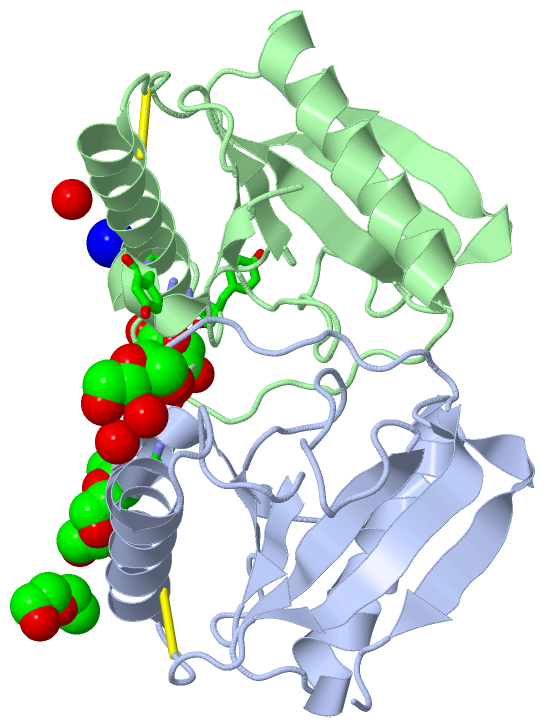
| | GO:0008745 | | N-acetylmuramoyl-L-alanine amidase activity | | Catalysis of the hydrolysis of the link between N-acetylmuramoyl residues and L-amino acid residues in certain bacterial cell-wall glycopeptides. |
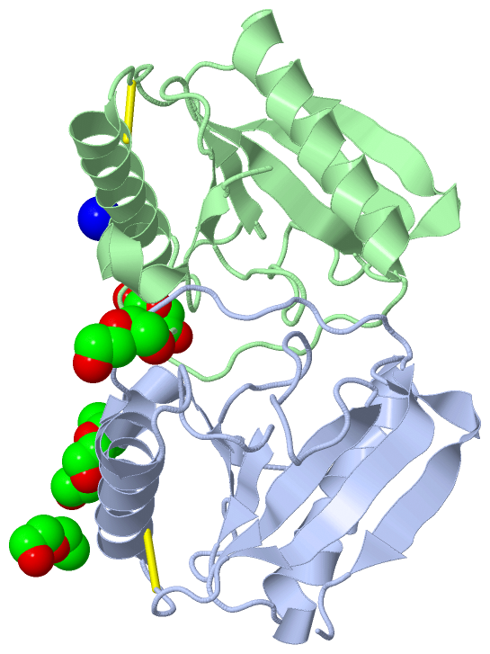
| | GO:0042834 | | peptidoglycan binding | | Interacting selectively and non-covalently, in a non-covalent manner, with peptidoglycan, any of a class of glycoconjugates found in bacterial cell walls. |
| | GO:0008270 | | zinc ion binding | | Interacting selectively and non-covalently with zinc (Zn) ions. |
| biological process |
|---|
| | GO:0019731 | | antibacterial humoral response | | An immune response against bacteria mediated through a body fluid. Examples of this process are the antibacterial humoral responses in Mus musculus and Drosophila melanogaster. |
| | GO:0006952 | | defense response | | Reactions, triggered in response to the presence of a foreign body or the occurrence of an injury, which result in restriction of damage to the organism attacked or prevention/recovery from the infection caused by the attack. |
| | GO:0098542 | | defense response to other organism | | Reactions triggered in response to the presence of another organism that act to protect the cell or organism from damage caused by that organism. |
| | GO:0008340 | | determination of adult lifespan | | The control of viability and duration in the adult phase of the life-cycle. |
| | GO:0002376 | | immune system process | | Any process involved in the development or functioning of the immune system, an organismal system for calibrated responses to potential internal or invasive threats. |
| | GO:0045087 | | innate immune response | | Innate immune responses are defense responses mediated by germline encoded components that directly recognize components of potential pathogens. |
| | GO:0046329 | | negative regulation of JNK cascade | | Any process that stops, prevents, or reduces the frequency, rate or extent of signal transduction mediated by the JNK cascade. |
| | GO:0009253 | | peptidoglycan catabolic process | | The chemical reactions and pathways resulting in the breakdown of peptidoglycans, any of a class of glycoconjugates found in bacterial cell walls. |
| | GO:0061057 | | peptidoglycan recognition protein signaling pathway | | A series of molecular signals initiated by binding of peptidoglycan to a receptor on the surface of the target cell and ending with regulation of a downstream cellular process. The main outcome of the Imd signaling is the production of antimicrobial peptides. |
| | GO:0009617 | | response to bacterium | | Any process that results in a change in state or activity of a cell or an organism (in terms of movement, secretion, enzyme production, gene expression, etc.) as a result of a stimulus from a bacterium. |
| cellular component |
|---|
| | GO:0031240 | | external side of cell outer membrane | | The side of the outer membrane that is opposite to the side that faces the periplasm of the cell. |
| | GO:0016021 | | integral component of membrane | | The component of a membrane consisting of the gene products and protein complexes having at least some part of their peptide sequence embedded in the hydrophobic region of the membrane. |
| | GO:0005887 | | integral component of plasma membrane | | The component of the plasma membrane consisting of the gene products and protein complexes having at least some part of their peptide sequence embedded in the hydrophobic region of the membrane. |
| | GO:0016020 | | membrane | | A lipid bilayer along with all the proteins and protein complexes embedded in it an attached to it. |
 Description