Show PDB file:   
         Plain Text   HTML   (compressed file size)
QuickSearch:   
by PDB,NDB,UniProt,PROSITE Code or Search Term(s)  
(-)Asymmetric Unit
(-)Asym. Unit - sites
(-)Biological Unit 1
(-)Biol. Unit 1 - sites
collapse expand < >
Image Asymmetric Unit
Asymmetric Unit  (Jmol Viewer)
Image Asym. Unit - sites
Asym. Unit - sites  (Jmol Viewer)
Image Biological Unit 1
Biological Unit 1  (Jmol Viewer)
Image Biol. Unit 1 - sites
Biol. Unit 1 - sites  (Jmol Viewer)

(-) Description

Title :  STRUCTURE AND METAL-LOADING OF A SOLUBLE PERIPLASM CUPRO-PROTEIN: CU-CUCA-CLOSED (SEMET)
 
Authors :  K. J. Waldron, S. J. Firbank, S. J. Dainty, M. Perez-Rama, S. Tottey, N. J
Date :  19 Jul 10  (Deposition) - 11 Aug 10  (Release) - 18 May 11  (Revision)
Method :  X-RAY DIFFRACTION
Resolution :  2.40
Chains :  Asym. Unit :  A
Biol. Unit 1:  A  (2x)
Keywords :  Metal Binding Protein, Cupin (Keyword Search: [Gene Ontology, PubMed, Web (Google))
 
Reference :  K. J. Waldron, S. J. Firbank, S. J. Dainty, M. Perez-Rama, S. Tottey, N. J. Robinson
Structure And Metal Loading Of A Soluble Periplasm Cuproprotein.
J. Biol. Chem. V. 285 32504 2010
PubMed-ID: 20702411  |  Reference-DOI: 10.1074/JBC.M110.153080

(-) Compounds

Molecule 1 - SLL1785 PROTEIN
    Atcc27184
    ChainsA
    EngineeredYES
    Expression SystemESCHERICHIA COLI
    Expression System PlasmidPET29A
    Expression System StrainB834(DE3)
    Expression System Taxid562
    Organism ScientificSYNECHOCYSTIS SP. PCC 6803
    Organism Taxid1148
    SynonymCUCA

 Structural Features

(-) Chains, Units

  1
Asymmetric Unit A
Biological Unit 1 (2x)A

Summary Information (see also Sequences/Alignments below)

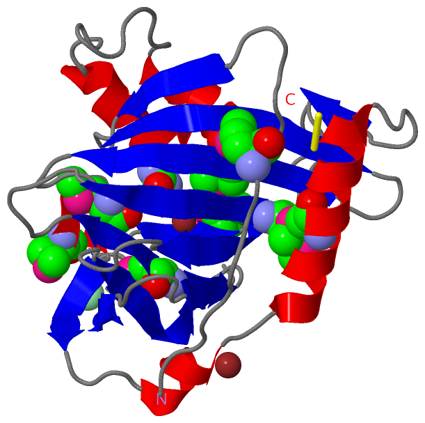
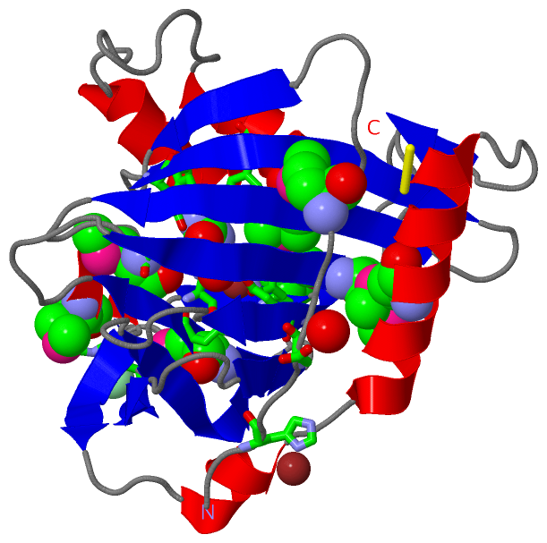
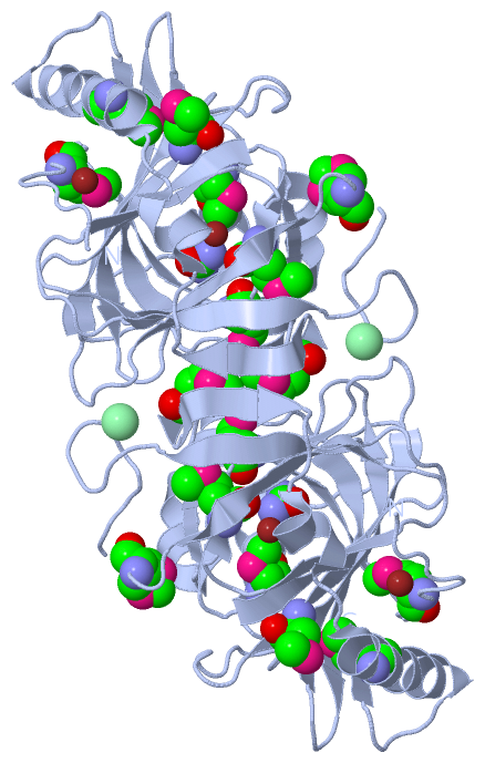
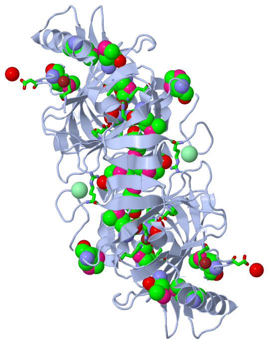
(-) Ligands, Modified Residues, Ions  (4, 12)

Asymmetric Unit (4, 12)
No.NameCountTypeFull Name
1CL1Ligand/IonCHLORIDE ION
2CU2Ligand/IonCOPPER (II) ION
3MSE8Mod. Amino AcidSELENOMETHIONINE
4URE1Ligand/IonUREA
Biological Unit 1 (2, 18)
No.NameCountTypeFull Name
1CL-1Ligand/IonCHLORIDE ION
2CU-1Ligand/IonCOPPER (II) ION
3MSE16Mod. Amino AcidSELENOMETHIONINE
4URE2Ligand/IonUREA

(-) Sites  (4, 4)

Asymmetric Unit (4, 4)
No.NameEvidenceResiduesDescription
1AC1SOFTWAREHIS A:88 , HIS A:90 , GLU A:95 , HIS A:149 , HOH A:2047BINDING SITE FOR RESIDUE CU A1269
2AC2SOFTWAREHIS A:36 , ASP A:39 , HOH A:2003 , HOH A:2005BINDING SITE FOR RESIDUE CU A1270
3AC3SOFTWAREGLU A:58 , PRO A:85 , PHE A:185 , PHE A:209 , HOH A:2047BINDING SITE FOR RESIDUE URE A1271
4AC4SOFTWAREARG A:127BINDING SITE FOR RESIDUE CL A1272

(-) SS Bonds  (1, 1)

Asymmetric Unit
No.Residues
1A:65 -A:267

(-) Cis Peptide Bonds  (0, 0)

(no "Cis Peptide Bond" information available for 2XL7)

 Sequence-Structure Mapping

(-) SAPs(SNPs)/Variants  (0, 0)

(no "SAP(SNP)/Variant" information available for 2XL7)

(-) PROSITE Motifs  (0, 0)

(no "PROSITE Motif" information available for 2XL7)

(-) Exons   (0, 0)

(no "Exon" information available for 2XL7)

(-) Sequences/Alignments

Asymmetric Unit
   Reformat: Number of residues per line =  ('0' or empty: single-line sequence representation)
  Number of residues per labelling interval =   
  UniProt sequence: complete  aligned part    
   Show mapping: SCOP domains CATH domains Pfam domains Secondary structure (by author)
SAPs(SNPs) PROSITE motifs Exons
(details for a mapped element are shown in a popup box when the mouse pointer rests over it)
Chain A from PDB  Type:PROTEIN  Length:235
 aligned with P73600_SYNY3 | P73600 from UniProtKB/TrEMBL  Length:268

    Alignment length:235
                                    43        53        63        73        83        93       103       113       123       133       143       153       163       173       183       193       203       213       223       233       243       253       263     
         P73600_SYNY3    34 EIHTFDDIPMPKLADPLLIYTPANEIFDIASCSAKDIGFAIAHAQIPPGGGPMPHIHYFINEWFWTPEGGIELFHSTKQYPNMDELPVVGGAGRGDLYSIQSEPKQLIYSPNHYMHGFVNPTDKTLPIVFVWMRNEVAPDFPYHDGGMREYFQAVGPRITDLNNLPELTNAQRAAFASEAPKYGINQSSYFMEYVNTISDKLPAQIAKLKNDKDLERMVEVIEAFNRGDKSVTCS 268
               SCOP domains ------------------------------------------------------------------------------------------------------------------------------------------------------------------------------------------------------------------------------------------- SCOP domains
               CATH domains ------------------------------------------------------------------------------------------------------------------------------------------------------------------------------------------------------------------------------------------- CATH domains
               Pfam domains ------------------------------------------------------------------------------------------------------------------------------------------------------------------------------------------------------------------------------------------- Pfam domains
         Sec.struct. author ................eeeee...eeeeeeeee....eeeeeeeee........eee..eeeeee......eeeeeeee...............eeeeeee.....eeee...eeeeee......eeeeeeeee............hhhhhhhhhh............hhhhhhhhhhhhhhh.eee..hhhh.eeeee...hhhhhh..hhhhhhhhhhhhhhhhh.....ee. Sec.struct. author
                 SAPs(SNPs) ------------------------------------------------------------------------------------------------------------------------------------------------------------------------------------------------------------------------------------------- SAPs(SNPs)
                    PROSITE ------------------------------------------------------------------------------------------------------------------------------------------------------------------------------------------------------------------------------------------- PROSITE
                 Transcript ------------------------------------------------------------------------------------------------------------------------------------------------------------------------------------------------------------------------------------------- Transcript
                 2xl7 A  34 EIHTFDDIPmPKLADPLLIYTPANEIFDIASCSAKDIGFAIAHAQIPPGGGPmPHIHYFINEWFWTPEGGIELFHSTKQYPNmDELPVVGGAGRGDLYSIQSEPKQLIYSPNHYmHGFVNPTDKTLPIVFVWmRNEVAPDFPYHDGGmREYFQAVGPRITDLNNLPELTNAQRAAFASEAPKYGINQSSYFmEYVNTISDKLPAQIAKLKNDKDLERmVEVIEAFNRGDKSVTCS 268
                                    43        53        63        73        83  |     93       103       113  |    123       133       143    |  153       163  |    173       183       193       203       213       223 |     233       243       253       263     
                                    43-MSE                                     86-MSE                       116-MSE                         148-MSE           166-MSE        181-MSE                                     225-MSE                   251-MSE             

   Legend:   → Mismatch (orange background)
  - → Gap (green background, '-', border residues have a numbering label)
    → Modified Residue (blue background, lower-case, 'x' indicates undefined single-letter code, labelled with number + name)
  x → Chemical Group (purple background, 'x', labelled with number + name, e.g. ACE or NH2)
  extra numbering lines below/above indicate numbering irregularities and modified residue names etc., number ends below/above '|'

 Classification and Annotation

(-) SCOP Domains  (0, 0)

(no "SCOP Domain" information available for 2XL7)

(-) CATH Domains  (0, 0)

(no "CATH Domain" information available for 2XL7)

(-) Pfam Domains  (0, 0)

(no "Pfam Domain" information available for 2XL7)

(-) Gene Ontology  (2, 2)

Asymmetric Unit(hide GO term definitions)
Chain A   (P73600_SYNY3 | P73600)
molecular function
    GO:0046872    metal ion binding    Interacting selectively and non-covalently with any metal ion.
cellular component
    GO:0030288    outer membrane-bounded periplasmic space    The region between the inner (cytoplasmic or plasma) membrane and outer membrane of organisms with two membranes such as Gram negative bacteria. These periplasmic spaces are relatively thick and contain a thin peptidoglycan layer (PGL), also referred to as a thin cell wall.

 Visualization

(-) Interactive Views

Asymmetric Unit
  Complete Structure
    Jena3D(integrated viewing of ligand, site, SAP, PROSITE, SCOP information)
    WebMol | AstexViewer[tm]@PDBe
(Java Applets, require no local installation except for Java; loading may be slow)
    STRAP
(Java WebStart application, automatic local installation, requires Java; full application with system access!)
    RasMol
(require local installation)
    Molscript (VRML)
(requires installation of a VRML viewer; select preferred view via VRML and generate a mono or stereo PDF format file)
 
  Ligands, Modified Residues, Ions
    CL  [ RasMol | Jena3D ]  +environment [ RasMol | Jena3D ]
    CU  [ RasMol | Jena3D ]  +environment [ RasMol | Jena3D ]
    MSE  [ RasMol | Jena3D ]  +environment [ RasMol | Jena3D ]
    URE  [ RasMol | Jena3D ]  +environment [ RasMol | Jena3D ]
 
  Sites
    AC1  [ RasMol ]  +environment [ RasMol ]
    AC2  [ RasMol ]  +environment [ RasMol ]
    AC3  [ RasMol ]  +environment [ RasMol ]
    AC4  [ RasMol ]  +environment [ RasMol ]
 
  Cis Peptide Bonds
(no "Cis Peptide Bonds" information available for 2xl7)
 
Biological Unit
  Complete Structure
    Biological Unit 1  [ Jena3D ]

(-) Still Images

Jmol
  protein: cartoon or spacefill or dots and stick; nucleic acid: cartoon and stick; ligands: spacefill; active site: stick
Molscript
  protein, nucleic acid: cartoon; ligands: spacefill; active site: ball and stick

 Databases and Analysis Tools

(-) Databases

Access by PDB/NDB ID
  2xl7
    Family and Domain InformationProDom | SYSTERS
    General Structural InformationGlycoscienceDB | MMDB | NDB | OCA | PDB | PDBe | PDBj | PDBsum | PDBWiki | PQS | PROTEOPEDIA
    Orientation in MembranesOPM
    Protein SurfaceSURFACE
    Secondary StructureDSSP (structure derived) | HSSP (homology derived)
    Structural GenomicsGeneCensus
    Structural NeighboursCE | VAST
    Structure ClassificationCATH | Dali | SCOP
    Validation and Original DataBMRB Data View | BMRB Restraints Grid | EDS | PROCHECK | RECOORD | WHAT_CHECK
 
Access by UniProt ID/Accession number
  P73600_SYNY3 | P73600
    Comparative Protein Structure ModelsModBase
    Genomic InformationEnsembl
    Protein-protein InteractionDIP
    Sequence, Family and Domain InformationInterPro | Pfam | SMART | UniProtKB/TrEMBL
 
Access by Enzyme Classificator   (EC Number)
  (no 'Enzyme Classificator' available)
    General Enzyme InformationBRENDA | EC-PDB | Enzyme | IntEnz
    PathwayKEGG | MetaCyc
 
Access by Disease Identifier   (MIM ID)
  (no 'MIM ID' available)
    Disease InformationOMIM
 
Access by GenAge ID
  (no 'GenAge ID' available)
    Age Related InformationGenAge

(-) Analysis Tools

Access by PDB/NDB ID
    Domain InformationXDom
    Interatomic Contacts of Structural UnitsCSU
    Ligand-protein ContactsLPC
    Protein CavitiescastP
    Sequence and Secondary StructurePDBCartoon
    Structure AlignmentSTRAP(Java WebStart application, automatic local installation, requires Java; full application with system access!)
    Structure and Sequence BrowserSTING
 
Access by UniProt ID/Accession number
  P73600_SYNY3 | P73600
    Protein Disorder PredictionDisEMBL | FoldIndex | GLOBPLOT (for more information see DisProt)

 Related Entries

(-) Entries Sharing at Least One Protein Chain (UniProt ID)

UniProtKB/TrEMBL
        P73600_SYNY3 | P736002xl9 2xla 2xlf 2xlg

(-) Related Entries Specified in the PDB File

2xl9 STRUCTURE AND METAL-LOADING OF A SOLUBLE PERIPLASM CUPRO-PROTEIN: ZN-CUCA-CLOSED (SEMET)
2xla STRUCTURE AND METAL-LOADING OF A SOLUBLE PERIPLASM CUPRO-PROTEIN: CU-CUCA-CLOSED
2xlf STRUCTURE AND METAL-LOADING OF A SOLUBLE PERIPLASM CUPRO-PROTEIN: APO-CUCA-CLOSED (SEMET)
2xlg STRUCTURE AND METAL-LOADING OF A SOLUBLE PERIPLASM CUPRO-PROTEIN: CU-CUCA-OPEN