|
|
|
|
 Description
|
|
Compounds
|
||||||||||||||||||||||||||||||||||||||||||||||||
Chains, Units
Summary Information (see also Sequences/Alignments below) |
Ligands, Modified Residues, Ions (3, 5)
Asymmetric Unit (3, 5)
|
Sites (3, 3)
Asymmetric Unit (3, 3)
|
SS Bonds (8, 8)
Asymmetric Unit
|
||||||||||||||||||||||||||||||||||||
Cis Peptide Bonds (0, 0)| (no "Cis Peptide Bond" information available for 2VLW) |
SAPs(SNPs)/Variants (0, 0)| (no "SAP(SNP)/Variant" information available for 2VLW) |
PROSITE Motifs (1, 2)
Asymmetric Unit (1, 2)
|
||||||||||||||||||||||||||||||||||||||||||||||||||||||||||||||||||||||||
Exons (0, 0)| (no "Exon" information available for 2VLW) |
Sequences/Alignments
Asymmetric UnitChain A from PDB Type:PROTEIN Length:65 aligned with 3SIM7_DENAN | Q8QGR0 from UniProtKB/Swiss-Prot Length:86 Alignment length:65 31 41 51 61 71 81 3SIM7_DENAN 22 LTCVKSNSIWFPTSEDCPDGQNLCFKRWQYISPRMYDFTRGCAATCPKAEYRDVINCCGTDKCNK 86 SCOP domains ----------------------------------------------------------------- SCOP domains CATH domains 2vlwA00 A:1-65 CD59 CATH domains Pfam domains ----------------------------------------------------------------- Pfam domains SAPs(SNPs) ----------------------------------------------------------------- SAPs(SNPs) PROSITE ----------------------------------------SNAKE_TOXIN ---- PROSITE Transcript ----------------------------------------------------------------- Transcript 2vlw A 1 LTCVKSNSIWFPTSEDCPDGQNLCFKRWQYISPRMYDFTRGCAATCPKAEyRDVINCCGTDKCNK 65 10 20 30 40 50| 60 51-TYI Chain B from PDB Type:PROTEIN Length:65 aligned with 3SIM7_DENAN | Q8QGR0 from UniProtKB/Swiss-Prot Length:86 Alignment length:65 31 41 51 61 71 81 3SIM7_DENAN 22 LTCVKSNSIWFPTSEDCPDGQNLCFKRWQYISPRMYDFTRGCAATCPKAEYRDVINCCGTDKCNK 86 SCOP domains ----------------------------------------------------------------- SCOP domains CATH domains 2vlwB00 B:1-65 CD59 CATH domains Pfam domains ----------------------------------------------------------------- Pfam domains SAPs(SNPs) ----------------------------------------------------------------- SAPs(SNPs) PROSITE ----------------------------------------SNAKE_TOXIN ---- PROSITE Transcript ----------------------------------------------------------------- Transcript 2vlw B 1 LTCVKSNSIWFPTSEDCPDGQNLCFKRWQYISPRMYDFTRGCAATCPKAEyRDVINCCGTDKCNK 65 10 20 30 40 50| 60 51-TYI
|
||||||||||||||||||||
SCOP Domains (0, 0)| (no "SCOP Domain" information available for 2VLW) |
CATH Domains (1, 2)
Asymmetric Unit
 |
Pfam Domains (0, 0)| (no "Pfam Domain" information available for 2VLW) |
Gene Ontology (5, 5)|
Asymmetric Unit(hide GO term definitions) Chain A,B (3SIM7_DENAN | Q8QGR0)
|
||||||||||||||||||||||||||||||||||||||||||||||||
Interactive Views
|
|||||||||||||||||||||||||||||||||||||||||||||||||||||||||||||||||||||||||||||||||||||||||||||||||||||||||||||||||||||||||||||||||||||||||||||||||||||||||||||||||||||||||
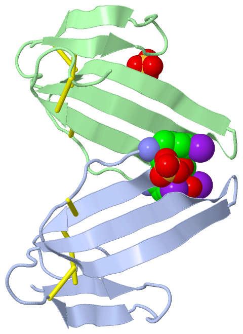
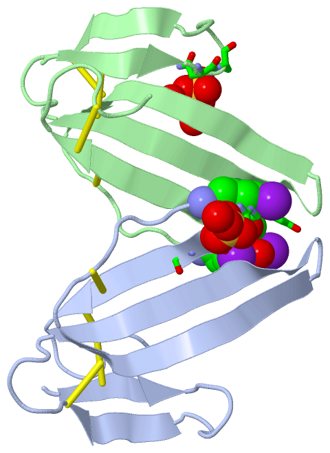
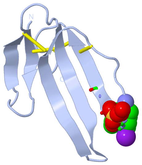
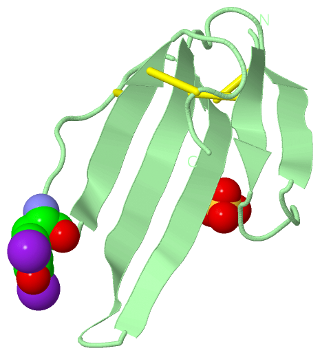
Still Images
|
||||||||||||||||
Databases
|
||||||||||||||||||||||||||||||||||||||||||||||||||||||||||||||||||||||||||||||||||||||||||||||||||||||||||||||||||||||||||||||||||||||||||||||||||||||||||||||||
Analysis Tools
|
|||||||||||||||||||||||||||||||||||||||||||||||||||||||||||||
Entries Sharing at Least One Protein Chain (UniProt ID)
Related Entries Specified in the PDB File
|
|