Show PDB file:   
         Plain Text   HTML   (compressed file size)
QuickSearch:   
by PDB,NDB,UniProt,PROSITE Code or Search Term(s)  
(-)NMR Structure - model 1
(-)NMR Structure - all models
collapse expand < >
Image NMR Structure - model 1
NMR Structure - model 1  (Jmol Viewer)
Image NMR Structure - all models
NMR Structure - all models  (Jmol Viewer)

(-) Description

Title :  SOLUTION STRUCTURE OF POLYTHEONAMIDE B
 
Authors :  N. Hamada, S. Matsunaga, M. Fujiwara, K. Fujjita, H. Hirota, R. Schmuck P. Guntert, N. Fusetani
Date :  03 Sep 09  (Deposition) - 15 Sep 10  (Release) - 24 Aug 11  (Revision)
Method :  SOLUTION NMR
Resolution :  NOT APPLICABLE
Chains :  NMR Structure  :  A  (20x)
NMR Structure *:  A  (1x)
Keywords :  Beta-Helix, Toxin (Keyword Search: [Gene Ontology, PubMed, Web (Google))
 
Reference :  T. Hamada, S. Matsunaga, M. Fujiwara, K. Fujita, H. Hirota, R. Schmucki, P. Guntert, N. Fusetani
Solution Structure Of Polytheonamide B, A Highly Cytotoxic Nonribosomal Polypeptide From Marine Sponge
J. Am. Chem. Soc. 2010
PubMed-ID: 20795624  |  Reference-DOI: 10.1021/JA104616Z

(-) Compounds

Molecule 1 - POLYTHEONAMIDE B
    ChainsA
    Organism ScientificTHEONELLA SWINHOEI
    Organism Taxid73791

 Structural Features

(-) Chains, Units

  1
NMR Structure (20x)A
NMR Structure * (1x)A

Summary Information (see also Sequences/Alignments below)

(-) Ligands, Modified Residues, Ions  (14, 28)

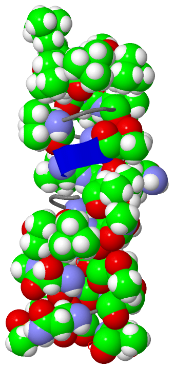
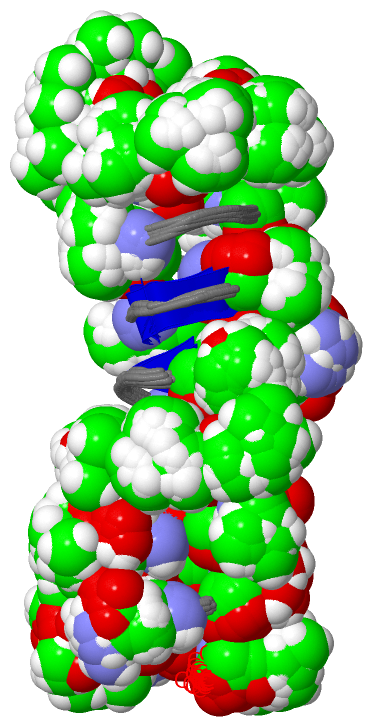
NMR Structure (14, 28)
No.NameCountTypeFull Name
12TL1Mod. Amino AcidD-ALLOTHREONINE
2DAL1Mod. Amino AcidD-ALANINE
3DHV2Mod. Amino Acid3-HYDROXY-D-VALINE
4DSG2Mod. Amino AcidD-ASPARAGINE
5DSN1Mod. Amino AcidD-SERINE
6HTN2Mod. Amino Acid(3R)-3-HYDROXY-N-METHYL-D-ASPARAGINE
7HVA1Mod. Amino Acid3-HYDROXY-L-VALINE
8I2M1Mod. Amino Acid3-METHYL-L-ALLOISOLEUCINE
9LMQ1Mod. Amino Acid(3S)-3-METHYL-L-GLUTAMINE
10M2S1Mod. Amino Acid3-{[(R)-METHYLSULFINYL]METHYL}-L-VALINE
11MHE1Mod. Amino Acid5,5-DIMETHYL-2-OXOHEXANOIC ACID
12MND6Mod. Amino AcidN-METHYL-D-ASPARAGINE
13TBG5Mod. Amino Acid3-METHYL-L-VALINE
14TDD3Mod. Amino Acid(2R)-2-AMINO-3,3-DIMETHYL-BUTANOIC ACID
NMR Structure * (14, 28)
No.NameCountTypeFull Name
12TL1Mod. Amino AcidD-ALLOTHREONINE
2DAL1Mod. Amino AcidD-ALANINE
3DHV2Mod. Amino Acid3-HYDROXY-D-VALINE
4DSG2Mod. Amino AcidD-ASPARAGINE
5DSN1Mod. Amino AcidD-SERINE
6HTN2Mod. Amino Acid(3R)-3-HYDROXY-N-METHYL-D-ASPARAGINE
7HVA1Mod. Amino Acid3-HYDROXY-L-VALINE
8I2M1Mod. Amino Acid3-METHYL-L-ALLOISOLEUCINE
9LMQ1Mod. Amino Acid(3S)-3-METHYL-L-GLUTAMINE
10M2S1Mod. Amino Acid3-{[(R)-METHYLSULFINYL]METHYL}-L-VALINE
11MHE1Mod. Amino Acid5,5-DIMETHYL-2-OXOHEXANOIC ACID
12MND6Mod. Amino AcidN-METHYL-D-ASPARAGINE
13TBG5Mod. Amino Acid3-METHYL-L-VALINE
14TDD3Mod. Amino Acid(2R)-2-AMINO-3,3-DIMETHYL-BUTANOIC ACID

(-) Sites  (0, 0)

(no "Site" information available for 2RQO)

(-) SS Bonds  (0, 0)

(no "SS Bond" information available for 2RQO)

(-) Cis Peptide Bonds  (0, 0)

(no "Cis Peptide Bond" information available for 2RQO)

 Sequence-Structure Mapping

(-) SAPs(SNPs)/Variants  (0, 0)

(no "SAP(SNP)/Variant" information available for 2RQO)

(-) PROSITE Motifs  (0, 0)

(no "PROSITE Motif" information available for 2RQO)

(-) Exons   (0, 0)

(no "Exon" information available for 2RQO)

(-) Sequences/Alignments

NMR Structure
   Reformat: Number of residues per line =  ('0' or empty: single-line sequence representation)
  Number of residues per labelling interval =   
  UniProt sequence: complete  aligned part    
   Show mapping: SCOP domains CATH domains Pfam domains Secondary structure (by author)
SAPs(SNPs) PROSITE motifs Exons
(details for a mapped element are shown in a popup box when the mouse pointer rests over it)
Chain A from PDB  Type:PROTEIN  Length:49
                                                                                
               SCOP domains ------------------------------------------------- SCOP domains
               CATH domains ------------------------------------------------- CATH domains
               Pfam domains ------------------------------------------------- Pfam domains
         Sec.struct. author ................................................. Sec.struct. author
                 SAPs(SNPs) ------------------------------------------------- SAPs(SNPs)
                    PROSITE ------------------------------------------------- PROSITE
                 Transcript ------------------------------------------------- Transcript
                  2rqo A  0 xGxGxxxxxxAGAxAxxGAGxxxxAGGxIxxxGxIxVxAxVxVxxxQxT 48
                            | | |||||9   | || 19||||   |29|| | | |39 | ||| | 
                            | | ||||||   | 16-HVA|||   | ||| | | | | | 44-M2S
                            0-MHE|||||   | |   20-TBG  | ||| | | | | | |45-DSG
                              2-I2M|||   | |    21-MND | ||| | | | | | |  47-2TL
                                4-TBG|   | |     22-LMQ| ||| | | | | | |     
                                 5-TDD   | |      23-DHV ||| | | | | | |     
                                  6-TBG  | |          27-MND | | | | | |     
                                   7-DAL | |            29-HTN | | | | |     
                                    8-TBG| |             30-TBG| | | | |     
                                     9-TDD |              31-DHV | | | |     

   Legend:   → Mismatch (orange background)
  - → Gap (green background, '-', border residues have a numbering label)
    → Modified Residue (blue background, lower-case, 'x' indicates undefined single-letter code, labelled with number + name)
  x → Chemical Group (purple background, 'x', labelled with number + name, e.g. ACE or NH2)
  extra numbering lines below/above indicate numbering irregularities and modified residue names etc., number ends below/above '|'

 Classification and Annotation

(-) SCOP Domains  (0, 0)

(no "SCOP Domain" information available for 2RQO)

(-) CATH Domains  (0, 0)

(no "CATH Domain" information available for 2RQO)

(-) Pfam Domains  (0, 0)

(no "Pfam Domain" information available for 2RQO)

(-) Gene Ontology  (0, 0)

NMR Structure(hide GO term definitions)
    (no "Gene Ontology" information available for 2RQO)

 Visualization

(-) Interactive Views

NMR Structure
  Complete Structure
    Jena3D(integrated viewing of ligand, site, SAP, PROSITE, SCOP information)
    WebMol | AstexViewer[tm]@PDBe
(Java Applets, require no local installation except for Java; loading may be slow)
    STRAP
(Java WebStart application, automatic local installation, requires Java; full application with system access!)
    RasMol
(require local installation)
    Molscript (VRML)
(requires installation of a VRML viewer; select preferred view via VRML and generate a mono or stereo PDF format file)
 
  Ligands, Modified Residues, Ions
    2TL  [ RasMol | Jena3D ]  +environment [ RasMol | Jena3D ]
    DAL  [ RasMol | Jena3D ]  +environment [ RasMol | Jena3D ]
    DHV  [ RasMol | Jena3D ]  +environment [ RasMol | Jena3D ]
    DSG  [ RasMol | Jena3D ]  +environment [ RasMol | Jena3D ]
    DSN  [ RasMol | Jena3D ]  +environment [ RasMol | Jena3D ]
    HTN  [ RasMol | Jena3D ]  +environment [ RasMol | Jena3D ]
    HVA  [ RasMol | Jena3D ]  +environment [ RasMol | Jena3D ]
    I2M  [ RasMol | Jena3D ]  +environment [ RasMol | Jena3D ]
    LMQ  [ RasMol | Jena3D ]  +environment [ RasMol | Jena3D ]
    M2S  [ RasMol | Jena3D ]  +environment [ RasMol | Jena3D ]
    MHE  [ RasMol | Jena3D ]  +environment [ RasMol | Jena3D ]
    MND  [ RasMol | Jena3D ]  +environment [ RasMol | Jena3D ]
    TBG  [ RasMol | Jena3D ]  +environment [ RasMol | Jena3D ]
    TDD  [ RasMol | Jena3D ]  +environment [ RasMol | Jena3D ]
 
  Sites
(no "Sites" information available for 2rqo)
 
  Cis Peptide Bonds
(no "Cis Peptide Bonds" information available for 2rqo)
 

(-) Still Images

Jmol
  protein: cartoon or spacefill or dots and stick; nucleic acid: cartoon and stick; ligands: spacefill; active site: stick
Molscript
  protein, nucleic acid: cartoon; ligands: spacefill; active site: ball and stick

 Databases and Analysis Tools

(-) Databases

Access by PDB/NDB ID
  2rqo
    Family and Domain InformationProDom | SYSTERS
    General Structural InformationGlycoscienceDB | MMDB | NDB | OCA | PDB | PDBe | PDBj | PDBsum | PDBWiki | PQS | PROTEOPEDIA
    Orientation in MembranesOPM
    Protein SurfaceSURFACE
    Secondary StructureDSSP (structure derived) | HSSP (homology derived)
    Structural GenomicsGeneCensus
    Structural NeighboursCE | VAST
    Structure ClassificationCATH | Dali | SCOP
    Validation and Original DataBMRB Data View | BMRB Restraints Grid | EDS | PROCHECK | RECOORD | WHAT_CHECK
 
Access by UniProt ID/Accession number
  (no 'UniProt ID/Accession number' available) |
    Comparative Protein Structure ModelsModBase
    Genomic InformationEnsembl
    Protein-protein InteractionDIP
    Sequence, Family and Domain InformationInterPro | Pfam | SMART | UniProt
 
Access by Enzyme Classificator   (EC Number)
  (no 'Enzyme Classificator' available)
    General Enzyme InformationBRENDA | EC-PDB | Enzyme | IntEnz
    PathwayKEGG | MetaCyc
 
Access by Disease Identifier   (MIM ID)
  (no 'MIM ID' available)
    Disease InformationOMIM
 
Access by GenAge ID
  (no 'GenAge ID' available)
    Age Related InformationGenAge

(-) Analysis Tools

Access by PDB/NDB ID
    Domain InformationXDom
    Interatomic Contacts of Structural UnitsCSU
    Ligand-protein ContactsLPC
    Protein CavitiescastP
    Sequence and Secondary StructurePDBCartoon
    Structure AlignmentSTRAP(Java WebStart application, automatic local installation, requires Java; full application with system access!)
    Structure and Sequence BrowserSTING
 
Access by UniProt ID/Accession number
  (no 'UniProt ID/Accession number' available) |
    Protein Disorder PredictionDisEMBL | FoldIndex | GLOBPLOT

 Related Entries

(-) Entries Sharing at Least One Protein Chain (UniProt ID)

(no "Entries Sharing at Least One Protein Chain" available for 2RQO)

(-) Related Entries Specified in the PDB File

(no "Related Entries Specified in the PDB File" available for 2RQO)