|
|
|
|
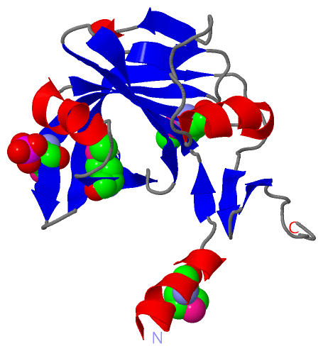
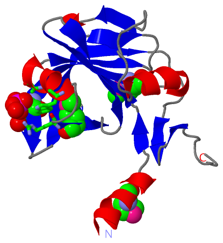
 Description
|
|
Compounds
|
||||||||||||||||||||||||||||||||||||||||||||||||||||
Chains, Units
Summary Information (see also Sequences/Alignments below) |
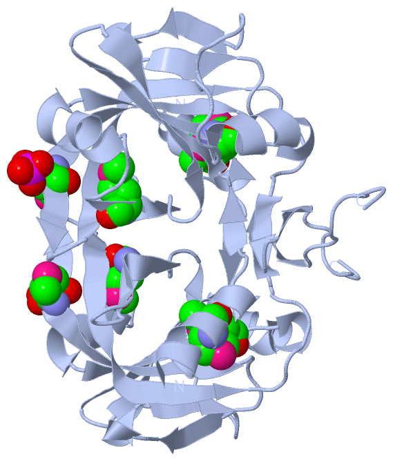
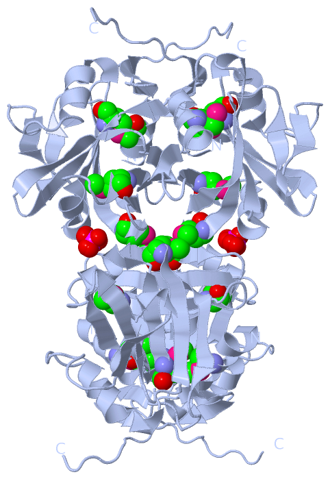
Ligands, Modified Residues, Ions (2, 5)| Asymmetric Unit (2, 5) Biological Unit 1 (2, 10) Biological Unit 2 (2, 20) |
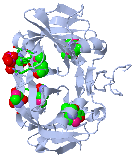
Sites (1, 1)
Asymmetric Unit (1, 1)
|
SS Bonds (0, 0)| (no "SS Bond" information available for 2QCK) |
Cis Peptide Bonds (1, 1)
Asymmetric Unit
|
||||||||
SAPs(SNPs)/Variants (0, 0)| (no "SAP(SNP)/Variant" information available for 2QCK) |
PROSITE Motifs (0, 0)| (no "PROSITE Motif" information available for 2QCK) |
Exons (0, 0)| (no "Exon" information available for 2QCK) |
Sequences/Alignments
Asymmetric UnitChain A from PDB Type:PROTEIN Length:159 aligned with A0JVA7_ARTS2 | A0JVA7 from UniProtKB/TrEMBL Length:166 Alignment length:159 17 27 37 47 57 67 77 87 97 107 117 127 137 147 157 A0JVA7_ARTS2 8 FEGTFKEMFRRHAAGVAIITVNYNGTPYGFTATSVASLSAQPPRFTFNMARSSSSWPAIANTTHIGVHMLGLDNQELADRFARTKNRFEGDHWELGPYEVPILKDVAGWLIGKIQMRLSFENNAVVVVEVVEGQVGEDGTPLLYHSGAYSQPVPLDYEI 166 SCOP domains d2qcka_ A: automated matches SCOP domains CATH domains 2qckA01 A:8-158 Electron Transport, Fmn-binding Protein; Chain A -------- CATH domains Pfam domains --------Flavin_Reduct-2qckA01 A:16-159 ------- Pfam domains SAPs(SNPs) --------------------------------------------------------------------------------------------------------------------------------------------------------------- SAPs(SNPs) PROSITE --------------------------------------------------------------------------------------------------------------------------------------------------------------- PROSITE Transcript --------------------------------------------------------------------------------------------------------------------------------------------------------------- Transcript 2qck A 8 FEGTFKEmFRRHAAGVAIITVNYNGTPYGFTATSVASLSAQPPRFTFNmARSSSSWPAIANTTHIGVHmLGLDNQELADRFARTKNRFEGDHWELGPYEVPILKDVAGWLIGKIQmRLSFENNAVVVVEVVEGQVGEDGTPLLYHSGAYSQPVPLDYEI 166 |17 27 37 47 57 67 77 87 97 107 117 | 127 137 147 157 15-MSE 56-MSE 76-MSE 123-MSE
|
||||||||||||||||||||
SCOP Domains (1, 1)
Asymmetric Unit
 |
CATH Domains (1, 1)
Asymmetric Unit
|
Pfam Domains (1, 1)
Asymmetric Unit
|
Gene Ontology (3, 3)|
Asymmetric Unit(hide GO term definitions) Chain A (A0JVA7_ARTS2 | A0JVA7)
|
||||||||||||||||||||||||||||||
Interactive Views
|
|||||||||||||||||||||||||||||||||||||||||||||||||||||||||||||||||||||||||||||||||||||||||||||||||||||||||||||||||||||||||||||||||||||||||||||||||||||
Still Images
|
||||||||||||||||
Databases
|
||||||||||||||||||||||||||||||||||||||||||||||||||||||||||||||||||||||||||||||||||||||||||||||||||||||||||||||||||||||||||||||||||||||||||||||||||||||||||||||||
Analysis Tools
|
|||||||||||||||||||||||||||||||||||||||||||||||||||||||||||||
Entries Sharing at Least One Protein Chain (UniProt ID)
Related Entries Specified in the PDB File
|
|