Show PDB file:   
         Plain Text   HTML   (compressed file size)
QuickSearch:   
by PDB,NDB,UniProt,PROSITE Code or Search Term(s)  
(-)Asym.Unit - manually
(-)Asymmetric Unit
(-)Asym. Unit - sites
(-)Biological Unit 1
(-)Biol. Unit 1 - sites
(-)Biological Unit 2
collapse expand < >
Image Asym.Unit - manually
Asym.Unit - manually  (Jmol Viewer)
Image Asymmetric Unit
Asymmetric Unit  (Jmol Viewer)
Image Asym. Unit - sites
Asym. Unit - sites  (Jmol Viewer)
Image Biological Unit 1
Biological Unit 1  (Jmol Viewer)
Image Biol. Unit 1 - sites
Biol. Unit 1 - sites  (Jmol Viewer)
Image Biological Unit 2
Biological Unit 2  (Jmol Viewer)

(-) Description

Title :  CRYSTAL STRUCTURE OF THE SECOND TETRAHEDRAL INTERMEDIATES OF SGPB AT PH 4.2
 
Authors :  T. W. Lee, M. N. G. James
Date :  14 Jun 07  (Deposition) - 11 Dec 07  (Release) - 13 Jul 11  (Revision)
Method :  X-RAY DIFFRACTION
Resolution :  1.18
Chains :  Asym. Unit :  E,I
Biol. Unit 1:  E,I  (2x)
Biol. Unit 2:  E,I  (1x)
Keywords :  Chymotrypsin-Type Serine Peptidase, Second Tetrahedral Intermediate, Tetrapeptide, Beta Barrels, Alpha Helix, Hydrolase (Keyword Search: [Gene Ontology, PubMed, Web (Google))
 
Reference :  T. W. Lee, M. N. James
1. 2A-Resolution Crystal Structures Reveal The Second Tetrahedral Intermediates Of Streptogrisin B (Sgpb).
Biochim. Biophys. Acta V. 1784 319 2008
PubMed-ID: 18157955  |  Reference-DOI: 10.1016/J.BBAPAP.2007.11.012

(-) Compounds

Molecule 1 - STREPTOGRISIN-B
    ChainsE
    EC Number3.4.21.81
    Organism ScientificSTREPTOMYCES GRISEUS
    Organism Taxid1911
    SynonymSERINE PROTEASE B, SGPB, PRONASE ENZYME B
 
Molecule 2 - 4-MER PEPTIDE DAIY
    ChainsI
    Organism ScientificSTREPTOMYCES GRISEUS
    Organism Taxid1911
    Other DetailsAUTO-PROTEOLYTIC PRODUCT

 Structural Features

(-) Chains, Units

  12
Asymmetric Unit EI
Biological Unit 1 (2x)EI
Biological Unit 2 (1x)EI

Summary Information (see also Sequences/Alignments below)

(-) Ligands, Modified Residues, Ions  (4, 9)

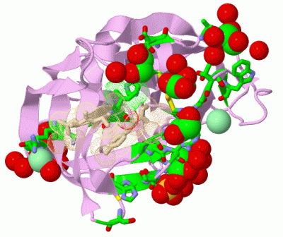
Asymmetric Unit (4, 9)
No.NameCountTypeFull Name
1CL2Ligand/IonCHLORIDE ION
2EDO4Ligand/Ion1,2-ETHANEDIOL
3GOL1Ligand/IonGLYCEROL
4SO42Ligand/IonSULFATE ION
Biological Unit 1 (3, 14)
No.NameCountTypeFull Name
1CL-1Ligand/IonCHLORIDE ION
2EDO8Ligand/Ion1,2-ETHANEDIOL
3GOL2Ligand/IonGLYCEROL
4SO44Ligand/IonSULFATE ION
Biological Unit 2 (3, 7)
No.NameCountTypeFull Name
1CL-1Ligand/IonCHLORIDE ION
2EDO4Ligand/Ion1,2-ETHANEDIOL
3GOL1Ligand/IonGLYCEROL
4SO42Ligand/IonSULFATE ION

(-) Sites  (9, 9)

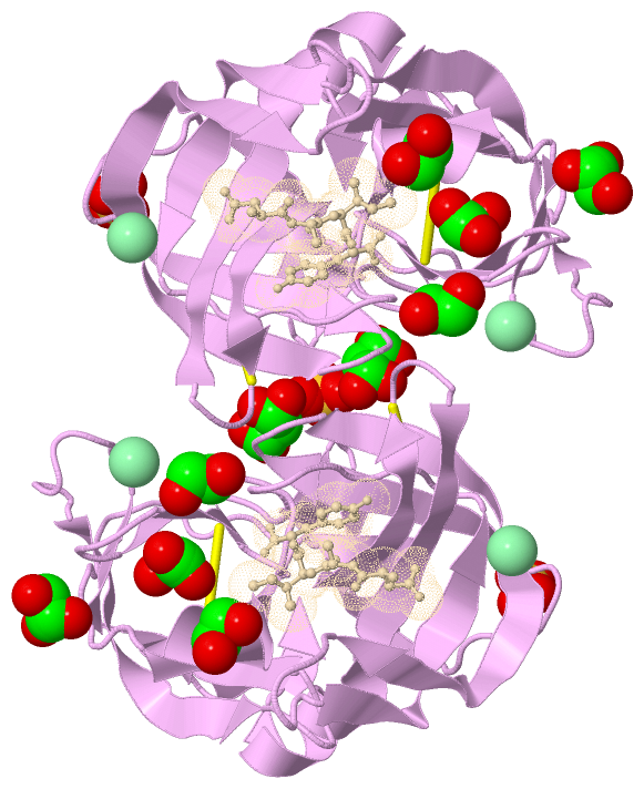
Asymmetric Unit (9, 9)
No.NameEvidenceResiduesDescription
1AC1SOFTWARETHR E:163 , ARG E:182 , HOH E:459 , HOH E:659BINDING SITE FOR RESIDUE CL E 411
2AC2SOFTWAREARG E:81BINDING SITE FOR RESIDUE CL E 412
3AC3SOFTWAREARG E:139 , GLY E:156 , THR E:157 , GOL E:401 , HOH E:655BINDING SITE FOR RESIDUE SO4 E 421
4AC4SOFTWAREARG E:208 , HOH E:463 , HOH E:489 , HOH E:564 , HOH E:592 , HOH E:651BINDING SITE FOR RESIDUE SO4 E 422
5AC5SOFTWARESER E:33 , THR E:39 , GLY E:40 , CYS E:42 , HOH E:514BINDING SITE FOR RESIDUE EDO E 431
6AC6SOFTWAREGLY E:40 , ARG E:41 , PRO E:192B , SER E:221BINDING SITE FOR RESIDUE EDO E 432
7AC7SOFTWAREHIS E:57 , ASP E:60 , HOH E:519 , HOH E:630 , HOH E:674 , ILE I:3BINDING SITE FOR RESIDUE EDO E 433
8AC8SOFTWARETHR E:64 , THR E:65 , TRP E:67 , VAL E:132 , HOH E:477 , HOH E:531 , HOH E:534 , HOH E:641BINDING SITE FOR RESIDUE EDO E 434
9AC9SOFTWAREGLY E:19 , ASP E:29 , ARG E:139 , GLY E:140 , SER E:141 , THR E:143 , GLY E:156 , THR E:157 , HIS E:158 , SO4 E:421 , HOH E:453 , HOH E:541 , HOH E:549 , HOH E:621BINDING SITE FOR RESIDUE GOL E 401

(-) SS Bonds  (2, 2)

Asymmetric Unit
No.Residues
1E:42 -E:58
2E:191 -E:220

(-) Cis Peptide Bonds  (1, 1)

Asymmetric Unit
No.Residues
1Phe E:94 -Pro E:99A

 Sequence-Structure Mapping

(-) SAPs(SNPs)/Variants  (0, 0)

(no "SAP(SNP)/Variant" information available for 2QA9)

(-) PROSITE Motifs  (2, 2)

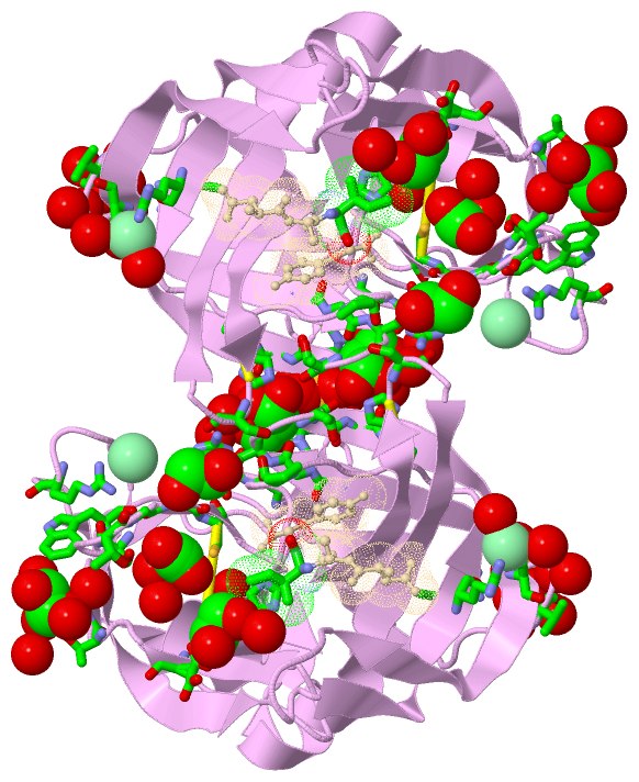
Asymmetric Unit (2, 2)
 PROSITEUniProtKBPDB
No.IDACDescriptionIDLocationCountLocation
1TRYPSIN_HISPS00134 Serine proteases, trypsin family, histidine active site.PRTB_STRGR143-148  1E:53-58
2TRYPSIN_SERPS00135 Serine proteases, trypsin family, serine active site.PRTB_STRGR249-260  1E:191-200
Biological Unit 1 (2, 4)
 PROSITEUniProtKBPDB
No.IDACDescriptionIDLocationCountLocation
1TRYPSIN_HISPS00134 Serine proteases, trypsin family, histidine active site.PRTB_STRGR143-148  2E:53-58
2TRYPSIN_SERPS00135 Serine proteases, trypsin family, serine active site.PRTB_STRGR249-260  2E:191-200
Biological Unit 2 (2, 2)
 PROSITEUniProtKBPDB
No.IDACDescriptionIDLocationCountLocation
1TRYPSIN_HISPS00134 Serine proteases, trypsin family, histidine active site.PRTB_STRGR143-148  1E:53-58
2TRYPSIN_SERPS00135 Serine proteases, trypsin family, serine active site.PRTB_STRGR249-260  1E:191-200

(-) Exons   (0, 0)

(no "Exon" information available for 2QA9)

(-) Sequences/Alignments

Asymmetric Unit
   Reformat: Number of residues per line =  ('0' or empty: single-line sequence representation)
  Number of residues per labelling interval =   
  UniProt sequence: complete  aligned part    
   Show mapping: SCOP domains CATH domains Pfam domains Secondary structure (by author)
SAPs(SNPs) PROSITE motifs Exons
(details for a mapped element are shown in a popup box when the mouse pointer rests over it)
Chain E from PDB  Type:PROTEIN  Length:185
 aligned with PRTB_STRGR | P00777 from UniProtKB/Swiss-Prot  Length:299

    Alignment length:185
                                   124       134       144       154       164       174       184       194       204       214       224       234       244       254       264       274       284       294     
          PRTB_STRGR    115 ISGGDAIYSSTGRCSLGFNVRSGSTYYFLTAGHCTDGATTWWANSARTTVLGTTSGSSFPNNDYGIVRYTNTTIPKDGTVGGQDITSAANATVGMAVTRRGSTTGTHSGSVTALNATVNYGGGDVVYGMIRTNVCAEPGDSGGPLYSGTRAIGLTSGGSGNCSSGGTTFFQPVTEALSAYGVSVY  299
               SCOP domains d2qa9e_ E: automated matches                                                                                                                                                              SCOP domains
               CATH domains 2qa9E01 E:16-116,E:231-242 Trypsin-like serine proteases                     2qa9E02 E:117-230 Trypsin-like serine proteases                                                2qa9E01       CATH domains
               Pfam domains Trypsin-2qa9E01 E:16-236                                                                                                                                                           ------ Pfam domains
         Sec.struct. author .....eee....eee..eeeee..eeeeeehhhhhh...eee.......eeeeeeeee.....eeeeee.........ee..ee..ee.......eeeeee...eeeeeeeeeeeeeee.....eeeeeeee...........eeee..eeeeeeeeeeee...eeeeeeeehhhhhhhhh.ee. Sec.struct. author
                 SAPs(SNPs) ----------------------------------------------------------------------------------------------------------------------------------------------------------------------------------------- SAPs(SNPs)
                    PROSITE ----------------------------TRYPSI----------------------------------------------------------------------------------------------------TRYPSIN_SER --------------------------------------- PROSITE
                 Transcript ----------------------------------------------------------------------------------------------------------------------------------------------------------------------------------------- Transcript
                2qa9 E   16 ISGGDAIYSSTGRCSLGFNVRSGSTYYFLTAGHCTDGATTWWANSARTTVLGTTSGSSFPNNDYGIVRYTNTTIPKDGTVGGQDITSAANATVGMAVTRRGSTTGTHSGSVTALNATVNYGGGDVVYGMIRTNVCAEPGDSGGPLYSGTRAIGLTSGGSGNCSSGGTTFFQPVTEALSAYGVSVY  242
                               ||   34|       48||||    54     || 65  ||    84      |99A       109       119       129       139   ||  161       171       181  ||  |194       208       218       228       237     
                              19|   34|       48A|||          60|    68|           91|||                                         143|                         184|  ||       202|                         235A       
                               29    39        48B||           62     78            93||                                          156                          190  ||        207                                    
                                                48C|                                 94|                                                                         192A|                                               
                                                 48D                                 99A                                                                          192B                                               

Chain I from PDB  Type:PROTEIN  Length:4
                                     
               SCOP domains ---- SCOP domains
               CATH domains ---- CATH domains
               Pfam domains ---- Pfam domains
         Sec.struct. author .ee. Sec.struct. author
                 SAPs(SNPs) ---- SAPs(SNPs)
                    PROSITE ---- PROSITE
                 Transcript ---- Transcript
                2qa9 I    1 DAIY    4

   Legend:   → Mismatch (orange background)
  - → Gap (green background, '-', border residues have a numbering label)
    → Modified Residue (blue background, lower-case, 'x' indicates undefined single-letter code, labelled with number + name)
  x → Chemical Group (purple background, 'x', labelled with number + name, e.g. ACE or NH2)
  extra numbering lines below/above indicate numbering irregularities and modified residue names etc., number ends below/above '|'

 Classification and Annotation

(-) SCOP Domains  (1, 1)

Asymmetric Unit

(-) CATH Domains  (1, 2)

Asymmetric Unit
(-)
Class: Mainly Beta (13760)

(-) Pfam Domains  (1, 1)

Asymmetric Unit

(-) Gene Ontology  (6, 6)

Asymmetric Unit(hide GO term definitions)
Chain E   (PRTB_STRGR | P00777)
molecular function
    GO:0016787    hydrolase activity    Catalysis of the hydrolysis of various bonds, e.g. C-O, C-N, C-C, phosphoric anhydride bonds, etc. Hydrolase is the systematic name for any enzyme of EC class 3.
    GO:0008233    peptidase activity    Catalysis of the hydrolysis of a peptide bond. A peptide bond is a covalent bond formed when the carbon atom from the carboxyl group of one amino acid shares electrons with the nitrogen atom from the amino group of a second amino acid.
    GO:0004252    serine-type endopeptidase activity    Catalysis of the hydrolysis of internal, alpha-peptide bonds in a polypeptide chain by a catalytic mechanism that involves a catalytic triad consisting of a serine nucleophile that is activated by a proton relay involving an acidic residue (e.g. aspartate or glutamate) and a basic residue (usually histidine).
    GO:0008236    serine-type peptidase activity    Catalysis of the hydrolysis of peptide bonds in a polypeptide chain by a catalytic mechanism that involves a catalytic triad consisting of a serine nucleophile that is activated by a proton relay involving an acidic residue (e.g. aspartate or glutamate) and a basic residue (usually histidine).
biological process
    GO:0006508    proteolysis    The hydrolysis of proteins into smaller polypeptides and/or amino acids by cleavage of their peptide bonds.
cellular component
    GO:0005576    extracellular region    The space external to the outermost structure of a cell. For cells without external protective or external encapsulating structures this refers to space outside of the plasma membrane. This term covers the host cell environment outside an intracellular parasite.

 Visualization

(-) Interactive Views

Asymmetric Unit
  Complete Structure
    Jena3D(integrated viewing of ligand, site, SAP, PROSITE, SCOP information)
    WebMol | AstexViewer[tm]@PDBe
(Java Applets, require no local installation except for Java; loading may be slow)
    STRAP
(Java WebStart application, automatic local installation, requires Java; full application with system access!)
    RasMol
(require local installation)
    Molscript (VRML)
(requires installation of a VRML viewer; select preferred view via VRML and generate a mono or stereo PDF format file)
 
  Ligands, Modified Residues, Ions
    CL  [ RasMol | Jena3D ]  +environment [ RasMol | Jena3D ]
    EDO  [ RasMol | Jena3D ]  +environment [ RasMol | Jena3D ]
    GOL  [ RasMol | Jena3D ]  +environment [ RasMol | Jena3D ]
    SO4  [ RasMol | Jena3D ]  +environment [ RasMol | Jena3D ]
 
  Sites
    AC1  [ RasMol ]  +environment [ RasMol ]
    AC2  [ RasMol ]  +environment [ RasMol ]
    AC3  [ RasMol ]  +environment [ RasMol ]
    AC4  [ RasMol ]  +environment [ RasMol ]
    AC5  [ RasMol ]  +environment [ RasMol ]
    AC6  [ RasMol ]  +environment [ RasMol ]
    AC7  [ RasMol ]  +environment [ RasMol ]
    AC8  [ RasMol ]  +environment [ RasMol ]
    AC9  [ RasMol ]  +environment [ RasMol ]
 
  Cis Peptide Bonds
    Phe E:94 - Pro E:99A  [ RasMol ]  
 
Biological Units
  Complete Structure
    Biological Unit 1  [ Jena3D ]
    Biological Unit 2  [ Jena3D ]

(-) Still Images

Jmol
  protein: cartoon or spacefill or dots and stick; nucleic acid: cartoon and stick; ligands: spacefill; active site: stick
Molscript
  protein, nucleic acid: cartoon; ligands: spacefill; active site: ball and stick

 Databases and Analysis Tools

(-) Databases

Access by PDB/NDB ID
  2qa9
    Family and Domain InformationProDom | SYSTERS
    General Structural InformationGlycoscienceDB | MMDB | NDB | OCA | PDB | PDBe | PDBj | PDBsum | PDBWiki | PQS | PROTEOPEDIA
    Orientation in MembranesOPM
    Protein SurfaceSURFACE
    Secondary StructureDSSP (structure derived) | HSSP (homology derived)
    Structural GenomicsGeneCensus
    Structural NeighboursCE | VAST
    Structure ClassificationCATH | Dali | SCOP
    Validation and Original DataBMRB Data View | BMRB Restraints Grid | EDS | PROCHECK | RECOORD | WHAT_CHECK
 
Access by UniProt ID/Accession number
  PRTB_STRGR | P00777
    Comparative Protein Structure ModelsModBase
    Genomic InformationEnsembl
    Protein-protein InteractionDIP
    Sequence, Family and Domain InformationInterPro | Pfam | SMART | UniProtKB/SwissProt
 
Access by Enzyme Classificator   (EC Number)
  3.4.21.81
    General Enzyme InformationBRENDA | EC-PDB | Enzyme | IntEnz
    PathwayKEGG | MetaCyc
 
Access by Disease Identifier   (MIM ID)
  (no 'MIM ID' available)
    Disease InformationOMIM
 
Access by GenAge ID
  (no 'GenAge ID' available)
    Age Related InformationGenAge

(-) Analysis Tools

Access by PDB/NDB ID
    Domain InformationXDom
    Interatomic Contacts of Structural UnitsCSU
    Ligand-protein ContactsLPC
    Protein CavitiescastP
    Sequence and Secondary StructurePDBCartoon
    Structure AlignmentSTRAP(Java WebStart application, automatic local installation, requires Java; full application with system access!)
    Structure and Sequence BrowserSTING
 
Access by UniProt ID/Accession number
  PRTB_STRGR | P00777
    Protein Disorder PredictionDisEMBL | FoldIndex | GLOBPLOT (for more information see DisProt)

 Related Entries

(-) Entries Sharing at Least One Protein Chain (UniProt ID)

UniProtKB/Swiss-Prot
        PRTB_STRGR | P007771cso 1ct0 1ct2 1ct4 1ds2 1sgd 1sge 1sgn 1sgp 1sgq 1sgr 1sgy 2gkv 2nu0 2nu1 2nu2 2nu3 2nu4 2qaa 2sgd 2sge 2sgf 2sgp 2sgq 3sgb 3sgq 4sgb

(-) Related Entries Specified in the PDB File

(no "Related Entries Specified in the PDB File" available for 2QA9)