Show PDB file:   
         Plain Text   HTML   (compressed file size)
QuickSearch:   
by PDB,NDB,UniProt,PROSITE Code or Search Term(s)  
(-)NMR Structure - model 1
(-)NMR Structure - all models
collapse expand < >
Image NMR Structure - model 1
NMR Structure - model 1  (Jmol Viewer)
Image NMR Structure - all models
NMR Structure - all models  (Jmol Viewer)

(-) Description

Title :  NMR STRCUTURE DETERMINATION OF THE PERIPLASMIC DOMAIN OF EXBD FROM E.COLI
 
Authors :  A. Garcia-Herrero, S. R. Peacock, P. S. Howard, H. J. Vogel
Date :  05 Apr 07  (Deposition) - 23 Oct 07  (Release) - 13 Jul 11  (Revision)
Method :  SOLUTION NMR
Resolution :  NOT APPLICABLE
Chains :  NMR Structure  :  A  (19x)
NMR Structure *:  A  (1x)
Keywords :  Exbd, Tonb System, Proton Motive Force, Periplasmic Domain, Transport Protein (Keyword Search: [Gene Ontology, PubMed, Web (Google))
 
Reference :  A. Garcia-Herrero, R. S. Peacock, S. P. Howard, H. J. Vogel
The Solution Structure Of The Periplasmic Domain Of The Ton System Exbd Protein Reveals An Unexpected Structural Homology With Siderophore-Binding Proteins.
Mol. Microbiol. V. 66 872 2007
PubMed-ID: 17927700  |  Reference-DOI: 10.1111/J.1365-2958.2007.05957.X

(-) Compounds

Molecule 1 - BIOPOLYMER TRANSPORT EXBD PROTEIN
    ChainsA
    FragmentPERIPLASMIC DOMAIN (RESIDUES 44-141)
    Organism ScientificESCHERICHIA COLI
    Organism Taxid83333
    StrainK12

 Structural Features

(-) Chains, Units

  1
NMR Structure (19x)A
NMR Structure * (1x)A

Summary Information (see also Sequences/Alignments below)

(-) Ligands, Modified Residues, Ions  (0, 0)

(no "Ligand,Modified Residues,Ions" information available for 2PFU)

(-) Sites  (0, 0)

(no "Site" information available for 2PFU)

(-) SS Bonds  (0, 0)

(no "SS Bond" information available for 2PFU)

(-) Cis Peptide Bonds  (0, 0)

(no "Cis Peptide Bond" information available for 2PFU)

 Sequence-Structure Mapping

(-) SAPs(SNPs)/Variants  (0, 0)

(no "SAP(SNP)/Variant" information available for 2PFU)

(-) PROSITE Motifs  (0, 0)

(no "PROSITE Motif" information available for 2PFU)

(-) Exons   (0, 0)

(no "Exon" information available for 2PFU)

(-) Sequences/Alignments

NMR Structure
   Reformat: Number of residues per line =  ('0' or empty: single-line sequence representation)
  Number of residues per labelling interval =   
  UniProt sequence: complete  aligned part    
   Show mapping: SCOP domains CATH domains Pfam domains Secondary structure (by author)
SAPs(SNPs) PROSITE motifs Exons
(details for a mapped element are shown in a popup box when the mouse pointer rests over it)
Chain A from PDB  Type:PROTEIN  Length:99
 aligned with EXBD_ECOLI | P0ABV2 from UniProtKB/Swiss-Prot  Length:141

    Alignment length:99
                                    52        62        72        82        92       102       112       122       132         
           EXBD_ECOLI    43 VDVKVNLPASTSTPQPRPEKPVYLSVKADNSMFIGNDPVTDETMITALNALTEGKKDTTIFFRADKTVDYETLMKVMDTLHQAGYLKIGLVGEETAKAK 141
               SCOP domains --------------------------------------------------------------------------------------------------- SCOP domains
               CATH domains --------------------------------------------------------------------------------------------------- CATH domains
               Pfam domains -ExbD-2pfuA01 A:44-137                                                                         ---- Pfam domains
         Sec.struct. author ......................eeeee...eeee..eee...hhhhhhhhhh.......eeeee....hhhhhhhhhhhhhhh....ee.......... Sec.struct. author
                 SAPs(SNPs) --------------------------------------------------------------------------------------------------- SAPs(SNPs)
                    PROSITE --------------------------------------------------------------------------------------------------- PROSITE
                 Transcript --------------------------------------------------------------------------------------------------- Transcript
                 2pfu A  43 MDVKVNLPASTSTPQPRPEKPVYLSVKADNSMFIGNDPVTDETMITALNALTEGKKDTTIFFRADKTVDYETLMKVMDTLHQAGYLKIGLVGEETAKAK 141
                                    52        62        72        82        92       102       112       122       132         

   Legend:   → Mismatch (orange background)
  - → Gap (green background, '-', border residues have a numbering label)
    → Modified Residue (blue background, lower-case, 'x' indicates undefined single-letter code, labelled with number + name)
  x → Chemical Group (purple background, 'x', labelled with number + name, e.g. ACE or NH2)
  extra numbering lines below/above indicate numbering irregularities and modified residue names etc., number ends below/above '|'

 Classification and Annotation

(-) SCOP Domains  (0, 0)

(no "SCOP Domain" information available for 2PFU)

(-) CATH Domains  (0, 0)

(no "CATH Domain" information available for 2PFU)

(-) Pfam Domains  (1, 1)

NMR Structure

(-) Gene Ontology  (11, 11)

NMR Structure(hide GO term definitions)
Chain A   (EXBD_ECOLI | P0ABV2)
molecular function
    GO:0031992    energy transducer activity    The biological transducer activity that accepts energy and converts it to another form, often by transfer to another molecule within the cell.
    GO:0042802    identical protein binding    Interacting selectively and non-covalently with an identical protein or proteins.
    GO:0005515    protein binding    Interacting selectively and non-covalently with any protein or protein complex (a complex of two or more proteins that may include other nonprotein molecules).
    GO:0005215    transporter activity    Enables the directed movement of substances (such as macromolecules, small molecules, ions) into, out of or within a cell, or between cells.
biological process
    GO:0043213    bacteriocin transport    The directed movement of a bacteriocin into, out of or within a cell, or between cells, by means of some agent such as a transporter or pore. Bacteriocins are a group of antibiotics produced by bacteria and are encoded by a group of naturally occurring plasmids, e.g. Col E1. Bacteriocins are toxic to bacteria closely related to the bacteriocin producing strain.
    GO:0050821    protein stabilization    Any process involved in maintaining the structure and integrity of a protein and preventing it from degradation or aggregation.
    GO:0015031    protein transport    The directed movement of proteins into, out of or within a cell, or between cells, by means of some agent such as a transporter or pore.
    GO:0006810    transport    The directed movement of substances (such as macromolecules, small molecules, ions) or cellular components (such as complexes and organelles) into, out of or within a cell, or between cells, or within a multicellular organism by means of some agent such as a transporter, pore or motor protein.
cellular component
    GO:0016021    integral component of membrane    The component of a membrane consisting of the gene products and protein complexes having at least some part of their peptide sequence embedded in the hydrophobic region of the membrane.
    GO:0016020    membrane    A lipid bilayer along with all the proteins and protein complexes embedded in it an attached to it.
    GO:0005886    plasma membrane    The membrane surrounding a cell that separates the cell from its external environment. It consists of a phospholipid bilayer and associated proteins.
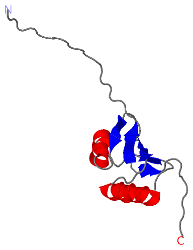
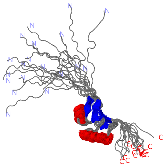
 Visualization

(-) Interactive Views

NMR Structure
  Complete Structure
    Jena3D(integrated viewing of ligand, site, SAP, PROSITE, SCOP information)
    WebMol | AstexViewer[tm]@PDBe
(Java Applets, require no local installation except for Java; loading may be slow)
    STRAP
(Java WebStart application, automatic local installation, requires Java; full application with system access!)
    RasMol
(require local installation)
    Molscript (VRML)
(requires installation of a VRML viewer; select preferred view via VRML and generate a mono or stereo PDF format file)
 
  Ligands, Modified Residues, Ions
(no "Ligands, Modified Residues, Ions" information available for 2pfu)
 
  Sites
(no "Sites" information available for 2pfu)
 
  Cis Peptide Bonds
(no "Cis Peptide Bonds" information available for 2pfu)
 

(-) Still Images

Jmol
  protein: cartoon or spacefill or dots and stick; nucleic acid: cartoon and stick; ligands: spacefill; active site: stick
Molscript
  protein, nucleic acid: cartoon; ligands: spacefill; active site: ball and stick

 Databases and Analysis Tools

(-) Databases

Access by PDB/NDB ID
  2pfu
    Family and Domain InformationProDom | SYSTERS
    General Structural InformationGlycoscienceDB | MMDB | NDB | OCA | PDB | PDBe | PDBj | PDBsum | PDBWiki | PQS | PROTEOPEDIA
    Orientation in MembranesOPM
    Protein SurfaceSURFACE
    Secondary StructureDSSP (structure derived) | HSSP (homology derived)
    Structural GenomicsGeneCensus
    Structural NeighboursCE | VAST
    Structure ClassificationCATH | Dali | SCOP
    Validation and Original DataBMRB Data View | BMRB Restraints Grid | EDS | PROCHECK | RECOORD | WHAT_CHECK
 
Access by UniProt ID/Accession number
  EXBD_ECOLI | P0ABV2
    Comparative Protein Structure ModelsModBase
    Genomic InformationEnsembl
    Protein-protein InteractionDIP
    Sequence, Family and Domain InformationInterPro | Pfam | SMART | UniProtKB/SwissProt
 
Access by Enzyme Classificator   (EC Number)
  (no 'Enzyme Classificator' available)
    General Enzyme InformationBRENDA | EC-PDB | Enzyme | IntEnz
    PathwayKEGG | MetaCyc
 
Access by Disease Identifier   (MIM ID)
  (no 'MIM ID' available)
    Disease InformationOMIM
 
Access by GenAge ID
  (no 'GenAge ID' available)
    Age Related InformationGenAge

(-) Analysis Tools

Access by PDB/NDB ID
    Domain InformationXDom
    Interatomic Contacts of Structural UnitsCSU
    Ligand-protein ContactsLPC
    Protein CavitiescastP
    Sequence and Secondary StructurePDBCartoon
    Structure AlignmentSTRAP(Java WebStart application, automatic local installation, requires Java; full application with system access!)
    Structure and Sequence BrowserSTING
 
Access by UniProt ID/Accession number
  EXBD_ECOLI | P0ABV2
    Protein Disorder PredictionDisEMBL | FoldIndex | GLOBPLOT (for more information see DisProt)

 Related Entries

(-) Entries Sharing at Least One Protein Chain (UniProt ID)

UniProtKB/Swiss-Prot
        EXBD_ECOLI | P0ABV25sv1

(-) Related Entries Specified in the PDB File

(no "Related Entries Specified in the PDB File" available for 2PFU)