Show PDB file:   
         Plain Text   HTML   (compressed file size)
QuickSearch:   
by PDB,NDB,UniProt,PROSITE Code or Search Term(s)  
(-)Biol.Unit 1 - manually
(-)Asym.Unit - manually
(-)Asymmetric Unit
(-)Asym. Unit - sites
(-)Biological Unit 1
(-)Biol. Unit 1 - sites
collapse expand < >
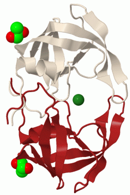
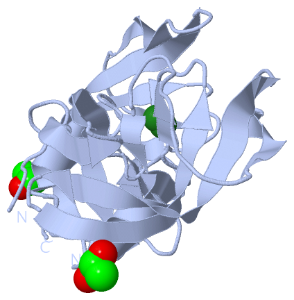
Image Biol.Unit 1 - manually
Biol.Unit 1 - manually  (Jmol Viewer)
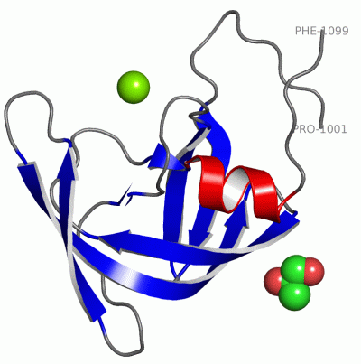
Image Asym.Unit - manually
Asym.Unit - manually  (Jmol Viewer)
Image Asymmetric Unit
Asymmetric Unit  (Jmol Viewer)
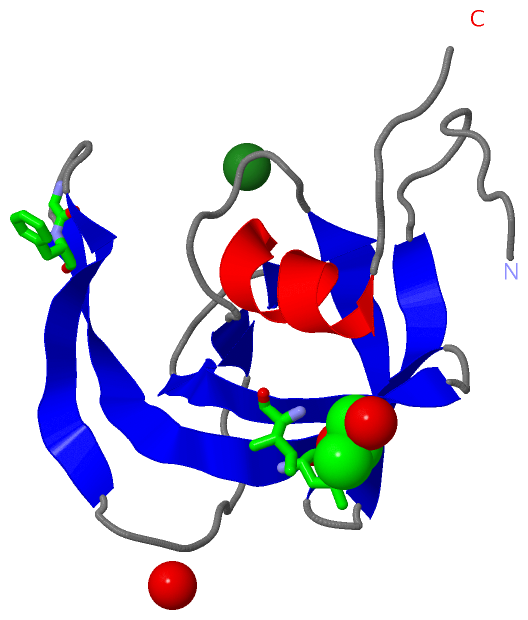
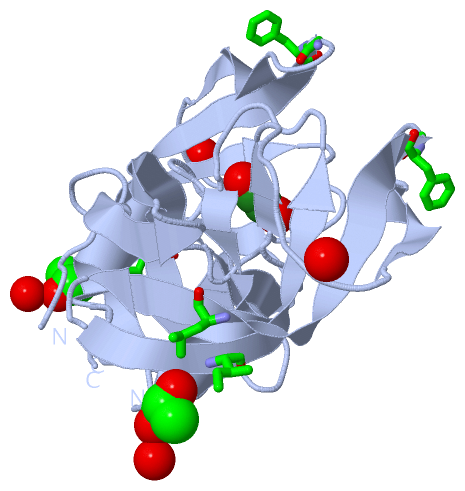
Image Asym. Unit - sites
Asym. Unit - sites  (Jmol Viewer)
Image Biological Unit 1
Biological Unit 1  (Jmol Viewer)
Image Biol. Unit 1 - sites
Biol. Unit 1 - sites  (Jmol Viewer)

(-) Description

Title :  APO WILD-TYPE HIV PROTEASE IN THE OPEN CONFORMATION
 
Authors :  H. Heaslet, R. Rosenfeld, M. J. Giffin, J. H. Elder, D. E. Mcree, C. D. Sto
Date :  29 Mar 07  (Deposition) - 26 Jun 07  (Release) - 13 Jul 11  (Revision)
Method :  X-RAY DIFFRACTION
Resolution :  1.40
Chains :  Asym. Unit :  A
Biol. Unit 1:  A  (2x)
Keywords :  Hiv Protease, Hydrolase (Keyword Search: [Gene Ontology, PubMed, Web (Google))
 
Reference :  H. Heaslet, R. Rosenfeld, M. Giffin, Y. C. Lin, K. Tam, B. E. Torbett, J. H. Elder, D. E. Mcree, C. D. Stout
Conformational Flexibility In The Flap Domains Of Ligand-Free Hiv Protease.
Acta Crystallogr. , Sect. D V. 63 866 2007
PubMed-ID: 17642513  |  Reference-DOI: 10.1107/S0907444907029125

(-) Compounds

Molecule 1 - PROTEASE
    ChainsA
    EC Number3.4.23.16
    EngineeredYES
    Expression SystemESCHERICHIA COLI
    Expression System PlasmidPLYS S
    Expression System StrainBL21.DE
    Expression System Taxid562
    Expression System Vector TypePLASMID
    GenePOL
    MutationYES
    Organism ScientificHUMAN IMMUNODEFICIENCY VIRUS 1
    Organism Taxid11676
    StrainBH10

 Structural Features

(-) Chains, Units

  1
Asymmetric Unit A
Biological Unit 1 (2x)A

Summary Information (see also Sequences/Alignments below)

(-) Ligands, Modified Residues, Ions  (2, 2)

Asymmetric Unit (2, 2)
No.NameCountTypeFull Name
1MG1Ligand/IonMAGNESIUM ION
2PGR1Ligand/IonR-1,2-PROPANEDIOL
Biological Unit 1 (1, 2)
No.NameCountTypeFull Name
1MG-1Ligand/IonMAGNESIUM ION
2PGR2Ligand/IonR-1,2-PROPANEDIOL

(-) Sites  (2, 2)

Asymmetric Unit (2, 2)
No.NameEvidenceResiduesDescription
1AC1SOFTWAREHOH A:1 , HOH A:50 , HOH A:62 , HOH A:85BINDING SITE FOR RESIDUE MG A 201
2AC2SOFTWAREHOH A:7 , HOH A:100 , GLY A:1052 , PHE A:1053 , LEU A:1063 , ILE A:1072BINDING SITE FOR RESIDUE PGR A 907

(-) SS Bonds  (0, 0)

(no "SS Bond" information available for 2PC0)

(-) Cis Peptide Bonds  (0, 0)

(no "Cis Peptide Bond" information available for 2PC0)

 Sequence-Structure Mapping

(-) SAPs(SNPs)/Variants  (0, 0)

(no "SAP(SNP)/Variant" information available for 2PC0)

(-) PROSITE Motifs  (0, 0)

(no "PROSITE Motif" information available for 2PC0)

(-) Exons   (0, 0)

(no "Exon" information available for 2PC0)

(-) Sequences/Alignments

Asymmetric Unit
   Reformat: Number of residues per line =  ('0' or empty: single-line sequence representation)
  Number of residues per labelling interval =   
  UniProt sequence: complete  aligned part    
   Show mapping: SCOP domains CATH domains Pfam domains Secondary structure (by author)
SAPs(SNPs) PROSITE motifs Exons
(details for a mapped element are shown in a popup box when the mouse pointer rests over it)
Chain A from PDB  Type:PROTEIN  Length:99
 aligned with A3FH86_9HIV1 | A3FH86 from UniProtKB/TrEMBL  Length:99

    Alignment length:99
                                    10        20        30        40        50        60        70        80        90         
        A3FH86_9HIV1      1 PQITLWQRPLVTIKIGGQLKEALLDTGADDTVLEEMNLPGRWKPKMIGGIGGFIKVRQYDQILIEICGHKAIGTVLVGPTPVNIIGRNLLTQIGCTLNF   99
               SCOP domains d2pc0a_ A: Human immunodeficiency virus type 1 protease                                             SCOP domains
               CATH domains 2pc0A00 A:1001-1099 Acid Proteases                                                                  CATH domains
               Pfam domains ---RVP-2pc0A01 A:1004-1098                                                                        - Pfam domains
         Sec.struct. author .........eeeeee..eeeeeee.......ee........eeeeeee....eeeeeeeeeeeeee..eeeeeeeee......eehhhhhhhh...... Sec.struct. author
                 SAPs(SNPs) --------------------------------------------------------------------------------------------------- SAPs(SNPs)
                    PROSITE --------------------------------------------------------------------------------------------------- PROSITE
                 Transcript --------------------------------------------------------------------------------------------------- Transcript
                2pc0 A 1001 PQITLWKRPLVTIKIGGQLKEALLDTGADDTVLEEMNLPGRWKPKMIGGIGGFIKVRQYDQILIEICGHKAIGTVLVGPTPVNIIGRNLLTQIGCTLNF 1099
                                  1010      1020      1030      1040      1050      1060      1070      1080      1090         

Chain A from PDB  Type:PROTEIN  Length:99
 aligned with Q903N5_9HIV1 | Q903N5 from UniProtKB/TrEMBL  Length:99

    Alignment length:99
                                    10        20        30        40        50        60        70        80        90         
        Q903N5_9HIV1      1 PQITLWRRPLVTIKIGGQLKEALLDTGADDTVLEEMNLPGRWKPKMIGGIGGFIKVRQYDQILIEICGHKAIGTVLVGPTPVNIIGRNLLTQIGCTLNF   99
               SCOP domains d2pc0a_ A: Human immunodeficiency virus type 1 protease                                             SCOP domains
               CATH domains 2pc0A00 A:1001-1099 Acid Proteases                                                                  CATH domains
               Pfam domains ---RVP-2pc0A01 A:1004-1098                                                                        - Pfam domains
         Sec.struct. author .........eeeeee..eeeeeee.......ee........eeeeeee....eeeeeeeeeeeeee..eeeeeeeee......eehhhhhhhh...... Sec.struct. author
                 SAPs(SNPs) --------------------------------------------------------------------------------------------------- SAPs(SNPs)
                    PROSITE --------------------------------------------------------------------------------------------------- PROSITE
                 Transcript --------------------------------------------------------------------------------------------------- Transcript
                2pc0 A 1001 PQITLWKRPLVTIKIGGQLKEALLDTGADDTVLEEMNLPGRWKPKMIGGIGGFIKVRQYDQILIEICGHKAIGTVLVGPTPVNIIGRNLLTQIGCTLNF 1099
                                  1010      1020      1030      1040      1050      1060      1070      1080      1090         

   Legend:   → Mismatch (orange background)
  - → Gap (green background, '-', border residues have a numbering label)
    → Modified Residue (blue background, lower-case, 'x' indicates undefined single-letter code, labelled with number + name)
  x → Chemical Group (purple background, 'x', labelled with number + name, e.g. ACE or NH2)
  extra numbering lines below/above indicate numbering irregularities and modified residue names etc., number ends below/above '|'

 Classification and Annotation

(-) SCOP Domains  (1, 1)

Asymmetric Unit

(-) CATH Domains  (1, 1)

Asymmetric Unit
(-)
Class: Mainly Beta (13760)

(-) Pfam Domains  (1, 1)

Asymmetric Unit

(-) Gene Ontology  (4, 8)

Asymmetric Unit(hide GO term definitions)
Chain A   (Q903N5_9HIV1 | Q903N5)
molecular function
    GO:0004190    aspartic-type endopeptidase activity    Catalysis of the hydrolysis of internal, alpha-peptide bonds in a polypeptide chain by a mechanism in which a water molecule bound by the side chains of aspartic residues at the active center acts as a nucleophile.
    GO:0016787    hydrolase activity    Catalysis of the hydrolysis of various bonds, e.g. C-O, C-N, C-C, phosphoric anhydride bonds, etc. Hydrolase is the systematic name for any enzyme of EC class 3.
    GO:0008233    peptidase activity    Catalysis of the hydrolysis of a peptide bond. A peptide bond is a covalent bond formed when the carbon atom from the carboxyl group of one amino acid shares electrons with the nitrogen atom from the amino group of a second amino acid.
biological process
    GO:0006508    proteolysis    The hydrolysis of proteins into smaller polypeptides and/or amino acids by cleavage of their peptide bonds.

Chain A   (A3FH86_9HIV1 | A3FH86)
molecular function
    GO:0004190    aspartic-type endopeptidase activity    Catalysis of the hydrolysis of internal, alpha-peptide bonds in a polypeptide chain by a mechanism in which a water molecule bound by the side chains of aspartic residues at the active center acts as a nucleophile.
    GO:0016787    hydrolase activity    Catalysis of the hydrolysis of various bonds, e.g. C-O, C-N, C-C, phosphoric anhydride bonds, etc. Hydrolase is the systematic name for any enzyme of EC class 3.
    GO:0008233    peptidase activity    Catalysis of the hydrolysis of a peptide bond. A peptide bond is a covalent bond formed when the carbon atom from the carboxyl group of one amino acid shares electrons with the nitrogen atom from the amino group of a second amino acid.
biological process
    GO:0006508    proteolysis    The hydrolysis of proteins into smaller polypeptides and/or amino acids by cleavage of their peptide bonds.

 Visualization

(-) Interactive Views

Asymmetric Unit
  Complete Structure
    Jena3D(integrated viewing of ligand, site, SAP, PROSITE, SCOP information)
    WebMol | AstexViewer[tm]@PDBe
(Java Applets, require no local installation except for Java; loading may be slow)
    STRAP
(Java WebStart application, automatic local installation, requires Java; full application with system access!)
    RasMol
(require local installation)
    Molscript (VRML)
(requires installation of a VRML viewer; select preferred view via VRML and generate a mono or stereo PDF format file)
 
  Ligands, Modified Residues, Ions
    MG  [ RasMol | Jena3D ]  +environment [ RasMol | Jena3D ]
    PGR  [ RasMol | Jena3D ]  +environment [ RasMol | Jena3D ]
 
  Sites
    AC1  [ RasMol ]  +environment [ RasMol ]
    AC2  [ RasMol ]  +environment [ RasMol ]
 
  Cis Peptide Bonds
(no "Cis Peptide Bonds" information available for 2pc0)
 
Biological Unit
  Complete Structure
    Biological Unit 1  [ Jena3D ]

(-) Still Images

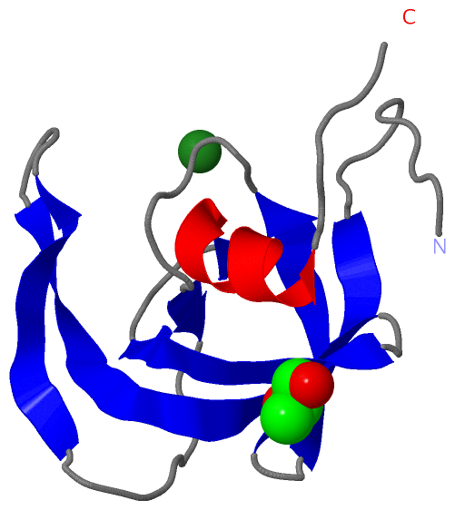
Jmol
  protein: cartoon or spacefill or dots and stick; nucleic acid: cartoon and stick; ligands: spacefill; active site: stick
Molscript
  protein, nucleic acid: cartoon; ligands: spacefill; active site: ball and stick

 Databases and Analysis Tools

(-) Databases

Access by PDB/NDB ID
  2pc0
    Family and Domain InformationProDom | SYSTERS
    General Structural InformationGlycoscienceDB | MMDB | NDB | OCA | PDB | PDBe | PDBj | PDBsum | PDBWiki | PQS | PROTEOPEDIA
    Orientation in MembranesOPM
    Protein SurfaceSURFACE
    Secondary StructureDSSP (structure derived) | HSSP (homology derived)
    Structural GenomicsGeneCensus
    Structural NeighboursCE | VAST
    Structure ClassificationCATH | Dali | SCOP
    Validation and Original DataBMRB Data View | BMRB Restraints Grid | EDS | PROCHECK | RECOORD | WHAT_CHECK
 
Access by UniProt ID/Accession number
  A3FH86_9HIV1 | A3FH86
    Comparative Protein Structure ModelsModBase
    Genomic InformationEnsembl
    Protein-protein InteractionDIP
    Sequence, Family and Domain InformationInterPro | Pfam | SMART | UniProtKB/SwissProt
  Q903N5_9HIV1 | Q903N5
    Comparative Protein Structure ModelsModBase
    Genomic InformationEnsembl
    Protein-protein InteractionDIP
    Sequence, Family and Domain InformationInterPro | Pfam | SMART | UniProtKB/TrEMBL
 
Access by Enzyme Classificator   (EC Number)
  3.4.23.16
    General Enzyme InformationBRENDA | EC-PDB | Enzyme | IntEnz
    PathwayKEGG | MetaCyc
 
Access by Disease Identifier   (MIM ID)
  (no 'MIM ID' available)
    Disease InformationOMIM
 
Access by GenAge ID
  (no 'GenAge ID' available)
    Age Related InformationGenAge

(-) Analysis Tools

Access by PDB/NDB ID
    Domain InformationXDom
    Interatomic Contacts of Structural UnitsCSU
    Ligand-protein ContactsLPC
    Protein CavitiescastP
    Sequence and Secondary StructurePDBCartoon
    Structure AlignmentSTRAP(Java WebStart application, automatic local installation, requires Java; full application with system access!)
    Structure and Sequence BrowserSTING
 
Access by UniProt ID/Accession number
  A3FH86_9HIV1 | A3FH86
    Protein Disorder PredictionDisEMBL | FoldIndex | GLOBPLOT (for more information see DisProt)
  Q903N5_9HIV1 | Q903N5
    Protein Disorder PredictionDisEMBL | FoldIndex | GLOBPLOT (for more information see DisProt)

 Related Entries

(-) Entries Sharing at Least One Protein Chain (UniProt ID)

UniProtKB/Swiss-Prot
        Q903N5_9HIV1 | Q903N53kf0 3kfn 3kfp 3kfr 3kfs 4e43
UniProtKB/TrEMBL
        Q903N5_9HIV1 | Q903N52hb4

(-) Related Entries Specified in the PDB File

2az8 WILD-TYPE HIV PROTEASE IN COMPLEX WITH TL-3.
2hb2 APO 6X MUTANT HIV PROTEASE IN THE CURLED CONFORMATION.
2hb4 APO WILD-TYPE HIV PROTEASE IN THE CURLED CONFORMATION.