Show PDB file:   
         Plain Text   HTML   (compressed file size)
QuickSearch:   
by PDB,NDB,UniProt,PROSITE Code or Search Term(s)  
(-)Asym./Biol. Unit
(-)Asym./Biol. Unit - sites
collapse expand < >
Image Asym./Biol. Unit
Asym./Biol. Unit  (Jmol Viewer)
Image Asym./Biol. Unit - sites
Asym./Biol. Unit - sites  (Jmol Viewer)

(-) Description

Title :  CRYSTAL STRUCTURE OF RHESUS ROTAVIRUS VP8* AT 100K
 
Authors :  H. Blanchard
Date :  09 Mar 07  (Deposition) - 11 Mar 08  (Release) - 13 Jul 11  (Revision)
Method :  X-RAY DIFFRACTION
Resolution :  1.56
Chains :  Asym./Biol. Unit :  A
Keywords :  Beta-Sandwich, Viral Protein (Keyword Search: [Gene Ontology, PubMed, Web (Google))
 
Reference :  M. J. Kraschnefski, A. Bugarcic, F. E. Fleming, X. Yu, M. Von Itzstein B. S. Coulson, H. Blanchard
Effects On Sialic Acid Recognition Of Amino Acid Mutations In The Carbohydrate-Binding Cleft Of The Rotavirus Spike Protein
Glycobiology V. 19 194 2009
PubMed-ID: 18974199  |  Reference-DOI: 10.1093/GLYCOB/CWN119

(-) Compounds

Molecule 1 - VP4
    ChainsA
    EngineeredYES
    Expression SystemESCHERICHIA COLI
    Expression System PlasmidPGEX-VP8*(64-224)
    Expression System StrainBL21 DE3
    Expression System Taxid469008
    Expression System Vector TypePLASMID
    FragmentVP8* DOMAIN (FRAGMENT 64-224)
    Organism ScientificRHESUS ROTAVIRUS
    Organism Taxid10969
    StrainRHESUS
    SynonymROTAVIRUS SPIKE PROTEIN

 Structural Features

(-) Chains, Units

  1
Asymmetric/Biological Unit A

Summary Information (see also Sequences/Alignments below)

(-) Ligands, Modified Residues, Ions  (3, 3)

Asymmetric/Biological Unit (3, 3)
No.NameCountTypeFull Name
1GOL1Ligand/IonGLYCEROL
2MNA1Ligand/Ion2-O-METHYL-5-N-ACETYL-ALPHA-D- NEURAMINIC ACID
3SO41Ligand/IonSULFATE ION

(-) Sites  (3, 3)

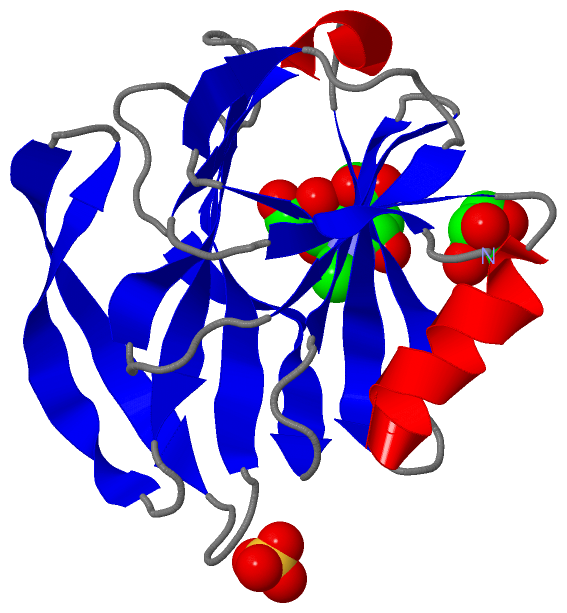
Asymmetric Unit (3, 3)
No.NameEvidenceResiduesDescription
1AC1SOFTWARESER A:134 , GLN A:135 , THR A:136 , HOH A:4102 , HOH A:4144 , HOH A:4153 , HOH A:4204 , HOH A:4213 , HOH A:4217BINDING SITE FOR RESIDUE SO4 A 3000
2AC2SOFTWAREARG A:101 , ASN A:111 , THR A:113 , ASN A:132 , ALA A:133 , TYR A:155 , LYS A:187 , TYR A:188 , TYR A:189 , SER A:190 , HOH A:4008 , HOH A:4108 , HOH A:4115 , HOH A:4157 , HOH A:4179 , HOH A:4195BINDING SITE FOR RESIDUE MNA A 1000
3AC3SOFTWAREMET A:168 , HIS A:170 , TYR A:175 , THR A:186 , ARG A:210 , GLU A:213BINDING SITE FOR RESIDUE GOL A 4000

(-) SS Bonds  (0, 0)

(no "SS Bond" information available for 2P3K)

(-) Cis Peptide Bonds  (2, 2)

Asymmetric/Biological Unit
No.Residues
1Gly A:67 -Pro A:68
2Thr A:181 -Pro A:182

 Sequence-Structure Mapping

(-) SAPs(SNPs)/Variants  (2, 2)

Asymmetric/Biological Unit (2, 2)
  dbSNPPDB
No.SourceVariant IDVariantUniProt IDStatusIDChainVariant
1UniProtVAR_VP4_ROTRH_001 *T146SVP4_ROTRH  ---  ---AT146S
2UniProtVAR_VP4_ROTRH_002 *A166GVP4_ROTRH  ---  ---AA166G
   * ID not provided by source

  SNP/SAP Summary Statistics (UniProtKB/Swiss-Prot)

(-) PROSITE Motifs  (0, 0)

(no "PROSITE Motif" information available for 2P3K)

(-) Exons   (0, 0)

(no "Exon" information available for 2P3K)

(-) Sequences/Alignments

Asymmetric/Biological Unit
   Reformat: Number of residues per line =  ('0' or empty: single-line sequence representation)
  Number of residues per labelling interval =   
  UniProt sequence: complete  aligned part    
   Show mapping: SCOP domains CATH domains Pfam domains Secondary structure (by author)
SAPs(SNPs) PROSITE motifs Exons
(details for a mapped element are shown in a popup box when the mouse pointer rests over it)
Chain A from PDB  Type:PROTEIN  Length:161
 aligned with VP4_ROTRH | P12473 from UniProtKB/Swiss-Prot  Length:776

    Alignment length:161
                                    73        83        93       103       113       123       133       143       153       163       173       183       193       203       213       223 
            VP4_ROTRH    64 VLDGPYQPTSFNPPVDYWMLLAPTAAGVVVEGTNNTDRWLATILVEPNVTSETRSYTLFGTQEQITIANASQTQWKFIDVVKTTQNGSYSQYGPLQSTPKLYAVMKHNGKIYTYNGETPNVTTKYYSTTNYDSVNMTAFCDFYIIPREEESTCTEYINNGL 224
               SCOP domains d2p3ka_ A: vp4 sialic acid binding domain                                                                                                                         SCOP domains
               CATH domains 2p3kA00 A:64-224  [code=2.60.120.200, no name defined]                                                                                                            CATH domains
               Pfam domains VP4_haemagglut-2p3kA01 A:64-224                                                                                                                                   Pfam domains
         Sec.struct. author ..eeee..eee.....eeeee.....eeeeeee.....eeeeeee...eeeeeeeeee..eeeeeeeee.....eeeeeeee.......eeeeeeee....eeeeee..eeeeeeee....eeeeee..hhhhheeee...eeeee..hhhhhhhhhhhh. Sec.struct. author
                 SAPs(SNPs) ----------------------------------------------------------------------------------S-------------------G---------------------------------------------------------- SAPs(SNPs)
                    PROSITE ----------------------------------------------------------------------------------------------------------------------------------------------------------------- PROSITE
                 Transcript ----------------------------------------------------------------------------------------------------------------------------------------------------------------- Transcript
                 2p3k A  64 VLDGPYQPTTFNPPVDYWMLLAPTAAGVVVEGTNNTDRWLATILVEPNVTSETRSYTLFGTQEQITIANASQTQWKFIDVVKTTQNGSYSQYGPLQSTPKLYAVMKHNGKIYTYNGETPNVTTKYYSTTNYDSVNMTAFCDFYIIPREEESTCTEYINNGL 224
                                    73        83        93       103       113       123       133       143       153       163       173       183       193       203       213       223 

   Legend:   → Mismatch (orange background)
  - → Gap (green background, '-', border residues have a numbering label)
    → Modified Residue (blue background, lower-case, 'x' indicates undefined single-letter code, labelled with number + name)
  x → Chemical Group (purple background, 'x', labelled with number + name, e.g. ACE or NH2)
  extra numbering lines below/above indicate numbering irregularities and modified residue names etc., number ends below/above '|'

 Classification and Annotation

(-) SCOP Domains  (1, 1)

Asymmetric/Biological Unit

(-) CATH Domains  (1, 1)

Asymmetric/Biological Unit
(-)
Class: Mainly Beta (13760)

(-) Pfam Domains  (1, 1)

Asymmetric/Biological Unit

(-) Gene Ontology  (10, 10)

Asymmetric/Biological Unit(hide GO term definitions)
Chain A   (VP4_ROTRH | P12473)
biological process
    GO:0039665    permeabilization of host organelle membrane involved in viral entry into host cell    Induction of organellar membrane permeabilization triggered by an interaction between the host membrane and a membrane-penetration protein associated with a viral capsid. Results in release of the virus contents from an organelle into the host cell cytoplasm.
    GO:0046718    viral entry into host cell    The process that occurs after viral attachment by which a virus, or viral nucleic acid, breaches the plasma membrane or cell envelope and enters the host cell. The process ends when the viral nucleic acid is released into the host cell cytoplasm.
    GO:0019058    viral life cycle    A set of processes which all viruses follow to ensure survival; includes attachment and entry of the virus particle, decoding of genome information, translation of viral mRNA by host ribosomes, genome replication, and assembly and release of viral particles containing the genome.
    GO:0016032    viral process    A multi-organism process in which a virus is a participant. The other participant is the host. Includes infection of a host cell, replication of the viral genome, and assembly of progeny virus particles. In some cases the viral genetic material may integrate into the host genome and only subsequently, under particular circumstances, 'complete' its life cycle.
    GO:0019062    virion attachment to host cell    The process by which a virion protein binds to molecules on the host cellular surface or host cell surface projection.
cellular component
    GO:0044165    host cell endoplasmic reticulum    The irregular network of unit membranes, visible only by electron microscopy, that occurs in the host cell cytoplasm of many eukaryotic cells. The membranes form a complex meshwork of tubular channels, which are often expanded into slitlike cavities called cisternae. The host ER takes two forms, rough (or granular), with ribosomes adhering to the outer surface, and smooth (with no ribosomes attached).
    GO:0044168    host cell rough endoplasmic reticulum    The irregular network of unit membranes, visible only by electron microscopy, that occurs in the host cell cytoplasm of many eukaryotic cells. The membranes form a complex meshwork of tubular channels, which are often expanded into slitlike cavities called cisternae. The host rough ER has ribosomes adhering to the outer surface.
    GO:0019028    viral capsid    The protein coat that surrounds the infective nucleic acid in some virus particles. It comprises numerous regularly arranged subunits, or capsomeres.
    GO:0039624    viral outer capsid    The outer layer of a double or triple concentric icosahedral capsid. Outer capsids are part of reoviridae and cystoviridae virions.
    GO:0019012    virion    The complete fully infectious extracellular virus particle.

 Visualization

(-) Interactive Views

Asymmetric/Biological Unit
  Complete Structure
    Jena3D(integrated viewing of ligand, site, SAP, PROSITE, SCOP information)
    WebMol | AstexViewer[tm]@PDBe
(Java Applets, require no local installation except for Java; loading may be slow)
    STRAP
(Java WebStart application, automatic local installation, requires Java; full application with system access!)
    RasMol
(require local installation)
    Molscript (VRML)
(requires installation of a VRML viewer; select preferred view via VRML and generate a mono or stereo PDF format file)
 
  Ligands, Modified Residues, Ions
    GOL  [ RasMol | Jena3D ]  +environment [ RasMol | Jena3D ]
    MNA  [ RasMol | Jena3D ]  +environment [ RasMol | Jena3D ]
    SO4  [ RasMol | Jena3D ]  +environment [ RasMol | Jena3D ]
 
  Sites
    AC1  [ RasMol ]  +environment [ RasMol ]
    AC2  [ RasMol ]  +environment [ RasMol ]
    AC3  [ RasMol ]  +environment [ RasMol ]
 
  Cis Peptide Bonds
    Gly A:67 - Pro A:68   [ RasMol ]  
    Thr A:181 - Pro A:182   [ RasMol ]  
 

(-) Still Images

Jmol
  protein: cartoon or spacefill or dots and stick; nucleic acid: cartoon and stick; ligands: spacefill; active site: stick
Molscript
  protein, nucleic acid: cartoon; ligands: spacefill; active site: ball and stick

 Databases and Analysis Tools

(-) Databases

Access by PDB/NDB ID
  2p3k
    Family and Domain InformationProDom | SYSTERS
    General Structural InformationGlycoscienceDB | MMDB | NDB | OCA | PDB | PDBe | PDBj | PDBsum | PDBWiki | PQS | PROTEOPEDIA
    Orientation in MembranesOPM
    Protein SurfaceSURFACE
    Secondary StructureDSSP (structure derived) | HSSP (homology derived)
    Structural GenomicsGeneCensus
    Structural NeighboursCE | VAST
    Structure ClassificationCATH | Dali | SCOP
    Validation and Original DataBMRB Data View | BMRB Restraints Grid | EDS | PROCHECK | RECOORD | WHAT_CHECK
 
Access by UniProt ID/Accession number
  VP4_ROTRH | P12473
    Comparative Protein Structure ModelsModBase
    Genomic InformationEnsembl
    Protein-protein InteractionDIP
    Sequence, Family and Domain InformationInterPro | Pfam | SMART | UniProtKB/SwissProt
 
Access by Enzyme Classificator   (EC Number)
  (no 'Enzyme Classificator' available)
    General Enzyme InformationBRENDA | EC-PDB | Enzyme | IntEnz
    PathwayKEGG | MetaCyc
 
Access by Disease Identifier   (MIM ID)
  (no 'MIM ID' available)
    Disease InformationOMIM
 
Access by GenAge ID
  (no 'GenAge ID' available)
    Age Related InformationGenAge

(-) Analysis Tools

Access by PDB/NDB ID
    Domain InformationXDom
    Interatomic Contacts of Structural UnitsCSU
    Ligand-protein ContactsLPC
    Protein CavitiescastP
    Sequence and Secondary StructurePDBCartoon
    Structure AlignmentSTRAP(Java WebStart application, automatic local installation, requires Java; full application with system access!)
    Structure and Sequence BrowserSTING
 
Access by UniProt ID/Accession number
  VP4_ROTRH | P12473
    Protein Disorder PredictionDisEMBL | FoldIndex | GLOBPLOT (for more information see DisProt)

 Related Entries

(-) Entries Sharing at Least One Protein Chain (UniProt ID)

UniProtKB/Swiss-Prot
        VP4_ROTRH | P124731kqr 1kri 1slq 2b4h 2b4i 2p3i 2p3j 3tb0

(-) Related Entries Specified in the PDB File

2p3i SAME NATIVE RHESUS ROTAVIRUS VP8* PROTEIN BUT STRUCTURE SOLVED AT 295K
2p3j RHESUS ROTAVIRUS VP8* WITH MUTATION ARG101 TO ALANINE. SOLVED AT 295K