|
|
|
|
 Description
|
|
Compounds
|
||||||||||||||||||||||||||||||||||||||||||||||||||||||||||||
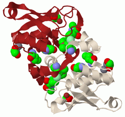
Chains, Units
Summary Information (see also Sequences/Alignments below) |
Ligands, Modified Residues, Ions (5, 10)| Asymmetric Unit (5, 10) Biological Unit 1 (2, 12) |
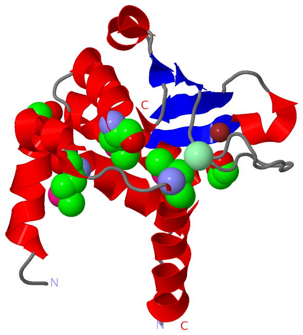
Sites (7, 7)
Asymmetric Unit (7, 7)
|
SS Bonds (0, 0)| (no "SS Bond" information available for 2OSD) |
Cis Peptide Bonds (1, 1)
Asymmetric Unit
|
||||||||
SAPs(SNPs)/Variants (0, 0)| (no "SAP(SNP)/Variant" information available for 2OSD) |
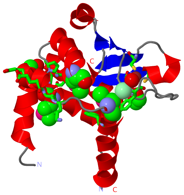
PROSITE Motifs (1, 1)
Asymmetric Unit (1, 1)
|
||||||||||||||||||||||||||||||||||||||||||||||||
Exons (0, 0)| (no "Exon" information available for 2OSD) |
Sequences/Alignments
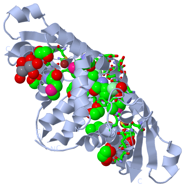
Asymmetric UnitChain A from PDB Type:PROTEIN Length:150 aligned with Y1460_METJA | Q58855 from UniProtKB/Swiss-Prot Length:162 Alignment length:152 14 24 34 44 54 64 74 84 94 104 114 124 134 144 154 Y1460_METJA 5 EKIFPDILEAIRNEEIIKESKKIPMPYFGLFALVIFDKVKELGSETSLYEIGEEFGKMLSPKNIEELKKIFKLMNFGDLEIDENKILLKNPPYKIKLSNPPYQWVSKEEPIHDFIAGILAGCLEEIFKKKFVVNEVECVSQGKDKCVFEVKE 156 SCOP domains d2osda1 A:5-156 Hypothetical protein MJ1 460 SCOP domains CATH domains 2osdA00 A:5-156 Trafficking protein part icle complex subunit 3 CATH domains Pfam domains -------------------------------------------------------------------------------------------V4R-2osdA01 A:96-156 Pfam domains SAPs(SNPs) -------------------------------------------------------------------------------------------------------------------------------------------------------- SAPs(SNPs) PROSITE --------------------------------------------------------------------------------------N6_MTAS----------------------------------------------------------- PROSITE Transcript -------------------------------------------------------------------------------------------------------------------------------------------------------- Transcript 2osd A 5 EKIFPDILEAIRNEEIIKESKKIPmPYFGLFALVIFDKVK--GSETSLYEIGEEFGKmLSPKNIEELKKIFKLmNFGDLEIDENKILLKNPPYKIKLSNPPYQWVSKEEPIHDFIAGILAGCLEEIFYYYFVVNEVECVSQGKDKCVFEVKE 156 14 24 | 34 44 | 54 |64 74 | 84 94 104 114 124 134 144 154 29-MSE 44 47 62-MSE 78-MSE
|
||||||||||||||||||||
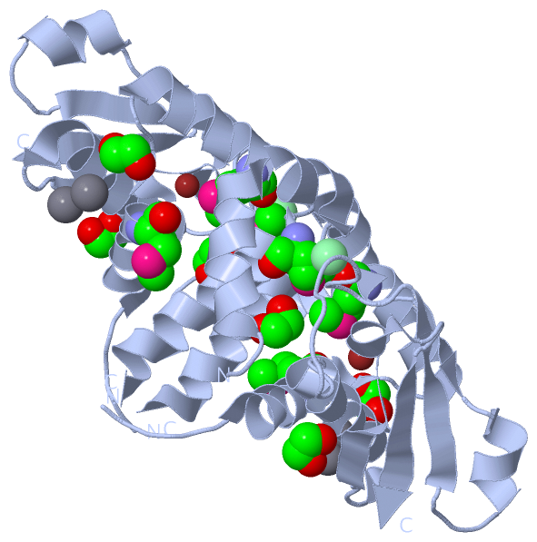
SCOP Domains (1, 1)
Asymmetric Unit
 |
CATH Domains (1, 1)
Asymmetric Unit
|
Pfam Domains (1, 1)
Asymmetric Unit
|
Gene Ontology (0, 0)|
Asymmetric Unit(hide GO term definitions)
(no "Gene Ontology" information available for 2OSD)
|
Interactive Views
|
|||||||||||||||||||||||||||||||||||||||||||||||||||||||||||||||||||||||||||||||||||||||||||||||||||||||||||||||||||||||||||||||||||||||||||||||||||||||||||||||||||||||||||||||||||||||||||||||||||||||||||||||
Still Images
|
||||||||||||||||
Databases
|
||||||||||||||||||||||||||||||||||||||||||||||||||||||||||||||||||||||||||||||||||||||||||||||||||||||||||||||||||||||||||||||||||||||||||||||||||||||||||||||||
Analysis Tools
|
|||||||||||||||||||||||||||||||||||||||||||||||||||||||||||||
Entries Sharing at Least One Protein Chain (UniProt ID)
Related Entries Specified in the PDB File
|
|