Show PDB file:   
         Plain Text   HTML   (compressed file size)
QuickSearch:   
by PDB,NDB,UniProt,PROSITE Code or Search Term(s)  
(-)Biol.Unit 1 - manually
(-)Asym.Unit - manually
(-)Asymmetric Unit
(-)Asym. Unit - sites
(-)Biological Unit 1
(-)Biol. Unit 1 - sites
collapse expand < >
Image Biol.Unit 1 - manually
Biol.Unit 1 - manually  (Jmol Viewer)
Image Asym.Unit - manually
Asym.Unit - manually  (Jmol Viewer)
Image Asymmetric Unit
Asymmetric Unit  (Jmol Viewer)
Image Asym. Unit - sites
Asym. Unit - sites  (Jmol Viewer)
Image Biological Unit 1
Biological Unit 1  (Jmol Viewer)
Image Biol. Unit 1 - sites
Biol. Unit 1 - sites  (Jmol Viewer)

(-) Description

Title :  CRYSTAL STRUCTURE OF A PEPTIDOGLYCAN GLYCOSYLTRANSFERASE FROM A CLASS A PBP: INSIGHT INTO BACTERIAL CELL WALL SYNTHESIS
 
Authors :  Y. Yuan, P. Sliz, S. Walker
Date :  31 Jan 07  (Deposition) - 13 Mar 07  (Release) - 13 Jul 11  (Revision)
Method :  X-RAY DIFFRACTION
Resolution :  2.10
Chains :  Asym. Unit :  A
Biol. Unit 1:  A  (2x)
Keywords :  Transferase (Keyword Search: [Gene Ontology, PubMed, Web (Google))
 
Reference :  Y. Yuan, D. Barrett, Y. Zhang, D. Kahne, P. Sliz, S. Walker
Crystal Structure Of A Peptidoglycan Glycosyltransferase Suggests A Model For Processive Glycan Chain Synthesis.
Proc. Natl. Acad. Sci. Usa V. 104 5348 2007
PubMed-ID: 17360321  |  Reference-DOI: 10.1073/PNAS.0701160104

(-) Compounds

Molecule 1 - PENICILLIN-BINDING PROTEIN 1A (PBP-1A) (PBP1A)
    ChainsA
    EC Number2.4.2.-
    EngineeredYES
    Expression SystemESCHERICHIA COLI BL21(DE3)
    Expression System PlasmidPET48(B)
    Expression System StrainBL21(DE3)
    Expression System Taxid469008
    Expression System Vector TypePLASMID
    FragmentGLYCOSYLTRANSFERASE DOMAIN
    GeneMRCA, PONA
    Organism ScientificAQUIFEX AEOLICUS
    Organism Taxid224324
    StrainVF5

 Structural Features

(-) Chains, Units

  1
Asymmetric Unit A
Biological Unit 1 (2x)A

Summary Information (see also Sequences/Alignments below)

(-) Ligands, Modified Residues, Ions  (2, 2)

Asymmetric Unit (2, 2)
No.NameCountTypeFull Name
1CPS1Ligand/Ion3-[(3-CHOLAMIDOPROPYL)DIMETHYLAMMONIO]-1-PROPANESULFONATE
2EPE1Ligand/Ion4-(2-HYDROXYETHYL)-1-PIPERAZINE ETHANESULFONIC ACID
Biological Unit 1 (2, 4)
No.NameCountTypeFull Name
1CPS2Ligand/Ion3-[(3-CHOLAMIDOPROPYL)DIMETHYLAMMONIO]-1-PROPANESULFONATE
2EPE2Ligand/Ion4-(2-HYDROXYETHYL)-1-PIPERAZINE ETHANESULFONIC ACID

(-) Sites  (2, 2)

Asymmetric Unit (2, 2)
No.NameEvidenceResiduesDescription
1AC1SOFTWAREALA A:81 , THR A:82 , ARG A:85 , ARG A:100 , VAL A:112 , LYS A:201 , ARG A:218 , ARG A:225BINDING SITE FOR RESIDUE EPE A 244
2AC2SOFTWAREILE A:93 , GLY A:114 , SER A:116 , LEU A:134 , ILE A:138 , ALA A:141 , LEU A:142 , ILE A:145BINDING SITE FOR RESIDUE CPS A 245

(-) SS Bonds  (0, 0)

(no "SS Bond" information available for 2OQO)

(-) Cis Peptide Bonds  (0, 0)

(no "Cis Peptide Bond" information available for 2OQO)

 Sequence-Structure Mapping

(-) SAPs(SNPs)/Variants  (0, 0)

(no "SAP(SNP)/Variant" information available for 2OQO)

(-) PROSITE Motifs  (0, 0)

(no "PROSITE Motif" information available for 2OQO)

(-) Exons   (0, 0)

(no "Exon" information available for 2OQO)

(-) Sequences/Alignments

Asymmetric Unit
   Reformat: Number of residues per line =  ('0' or empty: single-line sequence representation)
  Number of residues per labelling interval =   
  UniProt sequence: complete  aligned part    
   Show mapping: SCOP domains CATH domains Pfam domains Secondary structure (by author)
SAPs(SNPs) PROSITE motifs Exons
(details for a mapped element are shown in a popup box when the mouse pointer rests over it)
Chain A from PDB  Type:PROTEIN  Length:181
 aligned with PBPA_AQUAE | O66874 from UniProtKB/Swiss-Prot  Length:726

    Alignment length:187
                                    66        76        86        96       106       116       126       136       146       156       166       176       186       196       206       216       226       236       
           PBPA_AQUAE    57 TIGIQKRFYVSIDKIPEHVINAFVATEDRNFWHHFGIDPVAIVRAAIVNYRAGRIVQGGSTITQQLAKNLFLTRERTLERKIKEALLAIKIERTFDKKKIMELYLNQIYLGSGAYGVEAAAQVYFGKHVWELSLDEAALLAALPKAPAKYNPFYHPERALQRRNLVLKRMLEEGYITPEQYEEAVNK 243
               SCOP domains d2oqoa1 A:57-243 Penicillin-binding protein 1a, P      BP1a                                                                                                                                 SCOP domains
               CATH domains ------------------------------------------------------------------------------------------------------------------------------------------------------------------------------------------- CATH domains
               Pfam domains Transgly-2oqoA01 A:57-227                                                                                                                                                  ---------------- Pfam domains
         Sec.struct. author ..hhhhh...hhhhhhhhhhhhhhhhhh.........hhhhhhhhhh..------.....hhhhhhhhh.......hhhhhhhhhhhhhhhhhhhhhhhhhhhhhhhh.......hhhhhhhhhhh.hhhhhhhhhhhhhhhh..hhhhhh...hhhhhhhhhhhhhhhhhhh...hhhhhhhhhh. Sec.struct. author
                 SAPs(SNPs) ------------------------------------------------------------------------------------------------------------------------------------------------------------------------------------------- SAPs(SNPs)
                    PROSITE ------------------------------------------------------------------------------------------------------------------------------------------------------------------------------------------- PROSITE
                 Transcript ------------------------------------------------------------------------------------------------------------------------------------------------------------------------------------------- Transcript
                 2oqo A  57 TIGIQKRFYVSIDKIPEHVINAFVATEDRNFWHHFGIDPVAIVRAAIVN------VQGGSTITQQLAKNLFLTRERTLERKIKEALLAIKIERTFDKKKIMELYLNQIYLGSGAYGVEAAAQVYFGKHVWELSLDEAALLAALPKAPAKYNPFYHPERALQRRNLVLKRMLEEGYITPEQYEEAVNK 243
                                    66        76        86        96        |-     | 116       126       136       146       156       166       176       186       196       206       216       226       236       
                                                                          105    112                                                                                                                                   

   Legend:   → Mismatch (orange background)
  - → Gap (green background, '-', border residues have a numbering label)
    → Modified Residue (blue background, lower-case, 'x' indicates undefined single-letter code, labelled with number + name)
  x → Chemical Group (purple background, 'x', labelled with number + name, e.g. ACE or NH2)
  extra numbering lines below/above indicate numbering irregularities and modified residue names etc., number ends below/above '|'

 Classification and Annotation

(-) SCOP Domains  (1, 1)

Asymmetric Unit

(-) CATH Domains  (0, 0)

(no "CATH Domain" information available for 2OQO)

(-) Pfam Domains  (1, 1)

Asymmetric Unit

(-) Gene Ontology  (13, 13)

Asymmetric Unit(hide GO term definitions)
Chain A   (PBPA_AQUAE | O66874)
molecular function
    GO:0003824    catalytic activity    Catalysis of a biochemical reaction at physiological temperatures. In biologically catalyzed reactions, the reactants are known as substrates, and the catalysts are naturally occurring macromolecular substances known as enzymes. Enzymes possess specific binding sites for substrates, and are usually composed wholly or largely of protein, but RNA that has catalytic activity (ribozyme) is often also regarded as enzymatic.
    GO:0016787    hydrolase activity    Catalysis of the hydrolysis of various bonds, e.g. C-O, C-N, C-C, phosphoric anhydride bonds, etc. Hydrolase is the systematic name for any enzyme of EC class 3.
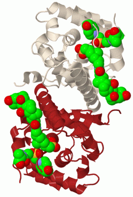
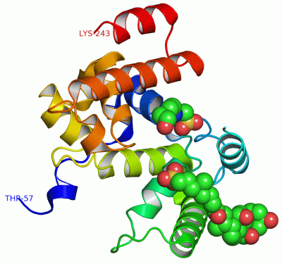
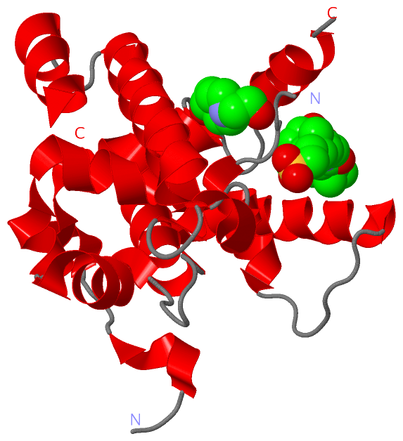
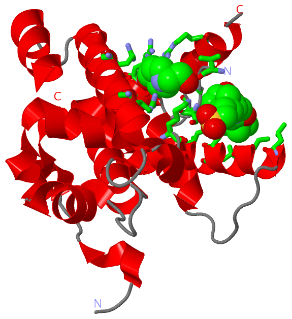
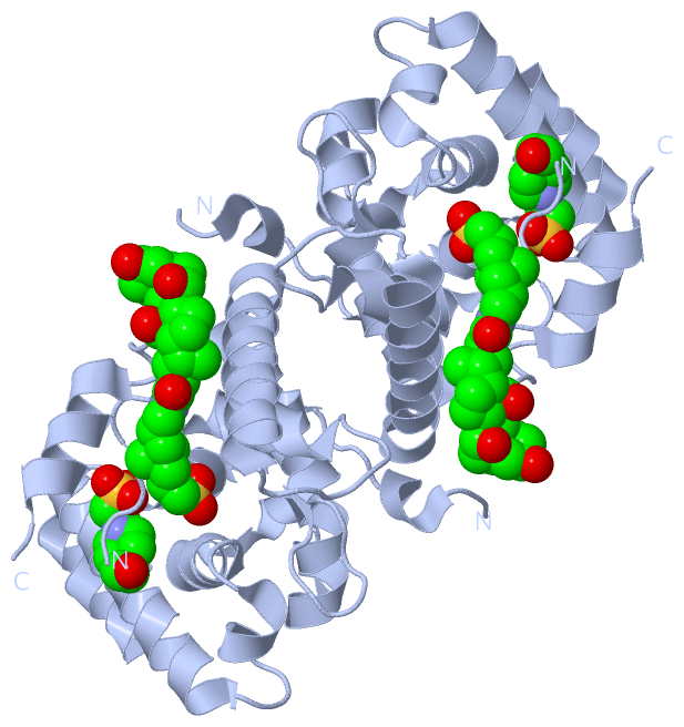
    GO:0008658    penicillin binding    Interacting selectively and non-covalently with penicillin, any antibiotic that contains the condensed beta-lactamthiazolidine ring system.
    GO:0016740    transferase activity    Catalysis of the transfer of a group, e.g. a methyl group, glycosyl group, acyl group, phosphorus-containing, or other groups, from one compound (generally regarded as the donor) to another compound (generally regarded as the acceptor). Transferase is the systematic name for any enzyme of EC class 2.
    GO:0016757    transferase activity, transferring glycosyl groups    Catalysis of the transfer of a glycosyl group from one compound (donor) to another (acceptor).
biological process
    GO:0071555    cell wall organization    A process that results in the assembly, arrangement of constituent parts, or disassembly of the cell wall, the rigid or semi-rigid envelope lying outside the cell membrane of plant, fungal and most prokaryotic cells, maintaining their shape and protecting them from osmotic lysis.
    GO:0008152    metabolic process    The chemical reactions and pathways, including anabolism and catabolism, by which living organisms transform chemical substances. Metabolic processes typically transform small molecules, but also include macromolecular processes such as DNA repair and replication, and protein synthesis and degradation.
    GO:0009252    peptidoglycan biosynthetic process    The chemical reactions and pathways resulting in the formation of peptidoglycans, any of a class of glycoconjugates found in bacterial cell walls.
    GO:0008360    regulation of cell shape    Any process that modulates the surface configuration of a cell.
    GO:0046677    response to antibiotic    Any process that results in a change in state or activity of a cell or an organism (in terms of movement, secretion, enzyme production, gene expression, etc.) as a result of an antibiotic stimulus. An antibiotic is a chemical substance produced by a microorganism which has the capacity to inhibit the growth of or to kill other microorganisms.
cellular component
    GO:0016021    integral component of membrane    The component of a membrane consisting of the gene products and protein complexes having at least some part of their peptide sequence embedded in the hydrophobic region of the membrane.
    GO:0016020    membrane    A lipid bilayer along with all the proteins and protein complexes embedded in it an attached to it.
    GO:0005886    plasma membrane    The membrane surrounding a cell that separates the cell from its external environment. It consists of a phospholipid bilayer and associated proteins.

 Visualization

(-) Interactive Views

Asymmetric Unit
  Complete Structure
    Jena3D(integrated viewing of ligand, site, SAP, PROSITE, SCOP information)
    WebMol | AstexViewer[tm]@PDBe
(Java Applets, require no local installation except for Java; loading may be slow)
    STRAP
(Java WebStart application, automatic local installation, requires Java; full application with system access!)
    RasMol
(require local installation)
    Molscript (VRML)
(requires installation of a VRML viewer; select preferred view via VRML and generate a mono or stereo PDF format file)
 
  Ligands, Modified Residues, Ions
    CPS  [ RasMol | Jena3D ]  +environment [ RasMol | Jena3D ]
    EPE  [ RasMol | Jena3D ]  +environment [ RasMol | Jena3D ]
 
  Sites
    AC1  [ RasMol ]  +environment [ RasMol ]
    AC2  [ RasMol ]  +environment [ RasMol ]
 
  Cis Peptide Bonds
(no "Cis Peptide Bonds" information available for 2oqo)
 
Biological Unit
  Complete Structure
    Biological Unit 1  [ Jena3D ]

(-) Still Images

Jmol
  protein: cartoon or spacefill or dots and stick; nucleic acid: cartoon and stick; ligands: spacefill; active site: stick
Molscript
  protein, nucleic acid: cartoon; ligands: spacefill; active site: ball and stick

 Databases and Analysis Tools

(-) Databases

Access by PDB/NDB ID
  2oqo
    Family and Domain InformationProDom | SYSTERS
    General Structural InformationGlycoscienceDB | MMDB | NDB | OCA | PDB | PDBe | PDBj | PDBsum | PDBWiki | PQS | PROTEOPEDIA
    Orientation in MembranesOPM
    Protein SurfaceSURFACE
    Secondary StructureDSSP (structure derived) | HSSP (homology derived)
    Structural GenomicsGeneCensus
    Structural NeighboursCE | VAST
    Structure ClassificationCATH | Dali | SCOP
    Validation and Original DataBMRB Data View | BMRB Restraints Grid | EDS | PROCHECK | RECOORD | WHAT_CHECK
 
Access by UniProt ID/Accession number
  PBPA_AQUAE | O66874
    Comparative Protein Structure ModelsModBase
    Genomic InformationEnsembl
    Protein-protein InteractionDIP
    Sequence, Family and Domain InformationInterPro | Pfam | SMART | UniProtKB/SwissProt
 
Access by Enzyme Classificator   (EC Number)
  2.4.2.-
    General Enzyme InformationBRENDA | EC-PDB | Enzyme | IntEnz
    PathwayKEGG | MetaCyc
 
Access by Disease Identifier   (MIM ID)
  (no 'MIM ID' available)
    Disease InformationOMIM
 
Access by GenAge ID
  (no 'GenAge ID' available)
    Age Related InformationGenAge

(-) Analysis Tools

Access by PDB/NDB ID
    Domain InformationXDom
    Interatomic Contacts of Structural UnitsCSU
    Ligand-protein ContactsLPC
    Protein CavitiescastP
    Sequence and Secondary StructurePDBCartoon
    Structure AlignmentSTRAP(Java WebStart application, automatic local installation, requires Java; full application with system access!)
    Structure and Sequence BrowserSTING
 
Access by UniProt ID/Accession number
  PBPA_AQUAE | O66874
    Protein Disorder PredictionDisEMBL | FoldIndex | GLOBPLOT (for more information see DisProt)

 Related Entries

(-) Entries Sharing at Least One Protein Chain (UniProt ID)

UniProtKB/Swiss-Prot
        PBPA_AQUAE | O668743d3h 3nb6 3nb7

(-) Related Entries Specified in the PDB File

(no "Related Entries Specified in the PDB File" available for 2OQO)