Show PDB file:   
         Plain Text   HTML   (compressed file size)
QuickSearch:   
by PDB,NDB,UniProt,PROSITE Code or Search Term(s)  
(-)Asym./Biol. Unit
collapse expand < >
Image Asym./Biol. Unit
Asym./Biol. Unit  (Jmol Viewer)

(-) Description

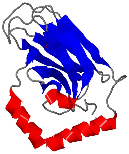
Title :  PEPTIDE-BINDING DOMAIN OF HEAT SHOCK 70 KDA PROTEIN D PRECURSOR FROM C.ELEGANS
 
Authors :  J. Osipiuk, E. Duggan, M. Gu, C. Voisine, R. I. Morimoto, A. Joachimiak, Center For Structural Genomics (Mcsg)
Date :  26 Jan 07  (Deposition) - 27 Feb 07  (Release) - 13 Jul 11  (Revision)
Method :  X-RAY DIFFRACTION
Resolution :  1.85
Chains :  Asym./Biol. Unit :  A
Keywords :  Hsp70/Peptide-Binding Domain, Structural Genomics, Apc90014. 13, Psi- 2, Protein Structure Initiative, Midwest Center For Structural Genomics, Mcsg, Peptide Binding Protein (Keyword Search: [Gene Ontology, PubMed, Web (Google)] )
 
Reference :  J. Osipiuk, E. Duggan, M. Gu, C. Voisine, R. I. Morimoto, A. Joachimiak
X-Ray Structure Of Peptide-Binding Domain Of Heat Shock 70 Kda Protein D Precursor From C. Elegans
To Be Published
PubMed: search

(-) Compounds

Molecule 1 - HEAT SHOCK 70 KDA PROTEIN D
    Chains: A
    Engineered: YES
    Expression System: ESCHERICHIA COLI BL21(DE3)
    Expression System Plasmid: PMCSG7
    Expression System Strain: BL21(DE3)
    Expression System Taxid: 469008
    Expression System Vector Type: PLASMID
    Fragment: HSP70 PEPTIDE-BINDING DOMAIN
    Gene: HSP-4, HSP70D
    Organism Scientific: CAENORHABDITIS ELEGANS
    Organism Taxid: 6239
    Strain: BRISTOL N2

 Structural Features

(-) Chains, Units

  1
Asymmetric/Biological Unit : A

Summary Information (see also Sequences/Alignments below)

(-) Ligands, Modified Residues, Ions  (0, 0)

(no "Ligand,Modified Residues,Ions" information available for 2OP6)

(-) Sites  (0, 0)

(no "Site" information available for 2OP6)

(-) SS Bonds  (0, 0)

(no "SS Bond" information available for 2OP6)

(-) Cis Peptide Bonds  (1, 1)

Asymmetric/Biological Unit
No.Residues
1Ile A:445 -Pro A:446

 Sequence-Structure Mapping

(-) SAPs(SNPs)/Variants  (0, 0)

(no "SAP(SNP)/Variant" information available for 2OP6)

(-) PROSITE Motifs  (0, 0)

(no "PROSITE Motif" information available for 2OP6)

(-) Exons   (0, 0)

(no "Exon" information available for 2OP6)

(-) Sequences/Alignments

Asymmetric/Biological Unit
   Reformat: Number of residues per line =  ('0' or empty: single-line sequence representation)
  Number of residues per labelling interval =   
  UniProt sequence: complete  aligned part    
   Show mapping: SCOP domains CATH domains Pfam domains Secondary structure (by author)
SAPs(SNPs) PROSITE motifs Exons
(details for a mapped element are shown in a popup box when the mouse pointer rests over it)
Chain A from PDB  Type:PROTEIN  Length:150
 aligned with HSP7D_CAEEL | P20163 from UniProtKB/Swiss-Prot  Length:657

    Alignment length:150
                                   428       438       448       458       468       478       488       498       508       518       528       538       548       558       568
          HSP7D_CAEEL   419 LDVNPLTLGIETVGGVMTKLIGRNTVIPTKKSQVFSTAADSQSAVSIVIYEGERPMVMDNHKLGNFDVTGIPPAPRGVPQIEVTFEIDVNGILHVSAEDKGTGNKNKLTITNDHNRLSPEDIERMINDADKFAADDQAQKEKVESRNELE 568
               SCOP domains ------------------------------------------------------------------------------------------------------------------------------------------------------ SCOP domains
               CATH domains 2op6A00 A:419-568 Substrate Binding Domain Of DNAk; Chain A, domain 1                                                                                  CATH domains
               Pfam domains -HSP70-2op6A01 A:420-568                                                                                                                               Pfam domains
         Sec.struct. author .......eeeee...eeeeee.......eeeeeeeee.......eeeeeee....hhhhheeeeeeee............eeeeeee.....eeeeeee.....eeeeee.......hhhhhhhhhhhhhhhhhhhhhhhhhh....ee. Sec.struct. author
                 SAPs(SNPs) ------------------------------------------------------------------------------------------------------------------------------------------------------ SAPs(SNPs)
                    PROSITE ------------------------------------------------------------------------------------------------------------------------------------------------------ PROSITE
                 Transcript ------------------------------------------------------------------------------------------------------------------------------------------------------ Transcript
                 2op6 A 419 ADVNPLTLGIETVGGVMTKLIGRNTVIPTKKSQVFSTAADSQSAVSIVIYEGERPMVMDNHKLGNFDVTGIPPAPRGVPQIEVTFEIDVNGILHVSAEDKGTGNKNKLTITNDHNRLSPEDIERMINDADKFAADDQAQKEKVESRNELE 568
                                   428       438       448       458       468       478       488       498       508       518       528       538       548       558       568

   Legend:   → Mismatch (orange background)
  - → Gap (green background, '-', border residues have a numbering label)
    → Modified Residue (blue background, lower-case, 'x' indicates undefined single-letter code, labelled with number + name)
  x → Chemical Group (purple background, 'x', labelled with number + name, e.g. ACE or NH2)
  extra numbering lines below/above indicate numbering irregularities and modified residue names etc., number ends below/above '|'

 Classification and Annotation

(-) SCOP Domains  (0, 0)

(no "SCOP Domain" information available for 2OP6)

(-) CATH Domains  (1, 1)

Asymmetric/Biological Unit
(-)
Class: Mainly Beta (13760)

(-) Pfam Domains  (1, 1)

Asymmetric/Biological Unit

(-) Gene Ontology  (9, 9)

Asymmetric/Biological Unit(hide GO term definitions)
Chain A   (HSP7D_CAEEL | P20163)
molecular function
    GO:0005524    ATP binding    Interacting selectively and non-covalently with ATP, adenosine 5'-triphosphate, a universally important coenzyme and enzyme regulator.
    GO:0001085    RNA polymerase II transcription factor binding    Interacting selectively and non-covalently with an RNA polymerase II transcription factor, any protein required to initiate or regulate transcription by RNA polymerase II.
    GO:0000166    nucleotide binding    Interacting selectively and non-covalently with a nucleotide, any compound consisting of a nucleoside that is esterified with (ortho)phosphate or an oligophosphate at any hydroxyl group on the ribose or deoxyribose.
biological process
    GO:0009792    embryo development ending in birth or egg hatching    The process whose specific outcome is the progression of an embryo over time, from zygote formation until the end of the embryonic life stage. The end of the embryonic life stage is organism-specific and may be somewhat arbitrary; for mammals it is usually considered to be birth, for insects the hatching of the first instar larva from the eggshell.
    GO:0030968    endoplasmic reticulum unfolded protein response    The series of molecular signals generated as a consequence of the presence of unfolded proteins in the endoplasmic reticulum (ER) or other ER-related stress; results in changes in the regulation of transcription and translation.
cellular component
    GO:0005783    endoplasmic reticulum    The irregular network of unit membranes, visible only by electron microscopy, that occurs in the cytoplasm of many eukaryotic cells. The membranes form a complex meshwork of tubular channels, which are often expanded into slitlike cavities called cisternae. The ER takes two forms, rough (or granular), with ribosomes adhering to the outer surface, and smooth (with no ribosomes attached).
    GO:0034663    endoplasmic reticulum chaperone complex    A protein complex that is located in the endoplasmic reticulum and is composed of chaperone proteins, including BiP, GRP94; CaBP1, protein disulfide isomerase (PDI), ERdj3, cyclophilin B, ERp72, GRP170, UDP-glucosyltransferase, and SDF2-L1.
    GO:0005788    endoplasmic reticulum lumen    The volume enclosed by the membranes of the endoplasmic reticulum.
    GO:0005791    rough endoplasmic reticulum    The rough (or granular) endoplasmic reticulum (ER) has ribosomes adhering to the outer surface; the ribosomes are the site of translation of the mRNA for those proteins which are either to be retained within the cisternae (ER-resident proteins), the proteins of the lysosomes, or the proteins destined for export from the cell. Glycoproteins undergo their initial glycosylation within the cisternae.

 Visualization

(-) Interactive Views

Asymmetric/Biological Unit
  Complete Structure
    Jena3D(integrated viewing of ligand, site, SAP, PROSITE, SCOP information)
    WebMol | AstexViewer[tm]@PDBe
(Java Applets, require no local installation except for Java; loading may be slow)
    STRAP
(Java WebStart application, automatic local installation, requires Java; full application with system access!)
    RasMol
(require local installation)
    Molscript (VRML)
(requires installation of a VRML viewer; select preferred view via VRML and generate a mono or stereo PDF format file)
 
  Ligands, Modified Residues, Ions
(no "Ligands, Modified Residues, Ions" information available for 2op6)
 
  Sites
(no "Sites" information available for 2op6)
 
  Cis Peptide Bonds
    Ile A:445 - Pro A:446   [ RasMol ]  
 

(-) Still Images

Jmol
  protein: cartoon or spacefill or dots and stick; nucleic acid: cartoon and stick; ligands: spacefill; active site: stick
Molscript
  protein, nucleic acid: cartoon; ligands: spacefill; active site: ball and stick

 Databases and Analysis Tools

(-) Databases

Access by PDB/NDB ID
  2op6
    Family and Domain Information: ProDom | SYSTERS
    General Structural Information: GlycoscienceDB | MMDB | NDB | OCA | PDB | PDBe | PDBj | PDBsum | PDBWiki | PQS | PROTEOPEDIA
    Orientation in Membranes: OPM
    Protein Surface: SURFACE
    Secondary Structure: DSSP (structure derived) | HSSP (homology derived)
    Structural Genomics: GeneCensus
    Structural Neighbours: CE | VAST
    Structure Classification: CATH | Dali | SCOP
    Validation and Original Data: BMRB Data View | BMRB Restraints Grid | EDS | PROCHECK | RECOORD | WHAT_CHECK
 
Access by UniProt ID/Accession number
  HSP7D_CAEEL | P20163
    Comparative Protein Structure Models: ModBase
    Genomic Information: Ensembl
    Protein-protein Interaction: DIP
    Sequence, Family and Domain Information: InterPro | Pfam | SMART | UniProtKB/SwissProt
 
Access by Enzyme Classificator   (EC Number)
  (no 'Enzyme Classificator' available)
    General Enzyme Information: BRENDA | EC-PDB | Enzyme | IntEnz
    Pathway: KEGG | MetaCyc
 
Access by Disease Identifier   (MIM ID)
  (no 'MIM ID' available)
    Disease Information: OMIM
 
Access by GenAge ID
  (no 'GenAge ID' available)
    Age Related Information: GenAge

(-) Analysis Tools

Access by PDB/NDB ID
    Domain Information: XDom
    Interatomic Contacts of Structural Units: CSU
    Ligand-protein Contacts: LPC
    Protein Cavities: castP
    Sequence and Secondary Structure: PDBCartoon
    Structure Alignment: STRAP(Java WebStart application, automatic local installation, requires Java; full application with system access!)
    Structure and Sequence Browser: STING
 
Access by UniProt ID/Accession number
  HSP7D_CAEEL | P20163
    Protein Disorder Prediction: DisEMBL | FoldIndex | GLOBPLOT (for more information see DisProt)

 Related Entries

(-) Entries Sharing at Least One Protein Chain (UniProt ID)

(no "Entries Sharing at Least One Protein Chain" available for 2OP6)

(-) Related Entries Specified in the PDB File

1dky SUBSTRATE BINDING DOMAIN OF DNAK IN COMPLEX WITH SUBSTRATE PEPTIDE
1yuw BOVINE HSC70(AA1-554)E213A/D214A MUTANT
7hsc HEAT SHOCK COGNATE-70 KD SUBSTRATE BINDING DOMAIN OBTAINED BY NMR