Show PDB file:   
         Plain Text   HTML   (compressed file size)
QuickSearch:   
by PDB,NDB,UniProt,PROSITE Code or Search Term(s)  
(-)NMR Structure - model 1
(-)NMR Structure - all models
collapse expand < >
Image NMR Structure - model 1
NMR Structure - model 1  (Jmol Viewer)
Image NMR Structure - all models
NMR Structure - all models  (Jmol Viewer)

(-) Description

Title :  NMR SOLUTION STRUCTURE OF OXIDIZED KPDSBA
 
Authors :  F. Kurth, K. Rimmer, L. Premkumar, B. Mohanty, W. Duprez, M. A. Halili, S. R. Shouldice, B. Heras, D. P. Fairlie, M. J. Scanlon, J. L. Martin
Date :  03 Aug 13  (Deposition) - 11 Dec 13  (Release) - 11 Dec 13  (Revision)
Method :  SOLUTION NMR
Resolution :  NOT APPLICABLE
Chains :  NMR Structure  :  A  (20x)
NMR Structure *:  A  (1x)
Keywords :  Oxidoreductase, Thioredoxin Domain (Keyword Search: [Gene Ontology, PubMed, Web (Google))
 
Reference :  F. Kurth, K. Rimmer, L. Premkumar, B. Mohanty, W. Duprez, M. A. Halili, S. R. Shouldice, B. Heras, D. P. Fairlie, M. J. Scanlon, J. L. Martin
Comparative Sequence, Structure And Redox Analyses Of Klebsiella Pneumoniae Dsba Show That Anti-Virulence Target Dsba Enzymes Fall Into Distinct Classes.
Plos One V. 8 80210 2013
PubMed-ID: 24244651  |  Reference-DOI: 10.1371/JOURNAL.PONE.0080210

(-) Compounds

Molecule 1
    ChainsA
    EngineeredYES
    Expression SystemESCHERICHIA COLI
    Expression System StrainROSETTA-2
    Expression System Taxid562
    Expression System VectorPMCSG-KPDSBA
    FragmentUNP RESIDUES 20-207
    GeneDSBA, KPK_5512
    Organism ScientificKLEBSIELLA PNEUMONIAE
    Organism Taxid507522
    Strain342

 Structural Features

(-) Chains, Units

  1
NMR Structure (20x)A
NMR Structure * (1x)A

Summary Information (see also Sequences/Alignments below)

(-) Ligands, Modified Residues, Ions  (0, 0)

(no "Ligand,Modified Residues,Ions" information available for 2MBS)

(-) Sites  (0, 0)

(no "Site" information available for 2MBS)
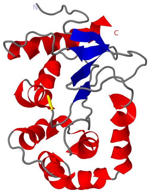
(-) SS Bonds  (1, 1)

NMR Structure
No.Residues
1A:30 -A:33

(-) Cis Peptide Bonds  (1, 20)

NMR Structure
No.ModelResidues
11, 2, 3, 4, 5, 6, 7, 8, 9, 10, 11, 12, 13, 14, 15, 16, 17, 18, 19, 20Val A:149 -Pro A:150

 Sequence-Structure Mapping

(-) SAPs(SNPs)/Variants  (0, 0)

(no "SAP(SNP)/Variant" information available for 2MBS)

(-) PROSITE Motifs  (0, 0)

(no "PROSITE Motif" information available for 2MBS)

(-) Exons   (0, 0)

(no "Exon" information available for 2MBS)

(-) Sequences/Alignments

NMR Structure
   Reformat: Number of residues per line =  ('0' or empty: single-line sequence representation)
  Number of residues per labelling interval =   
  UniProt sequence: complete  aligned part    
   Show mapping: SCOP domains CATH domains Pfam domains Secondary structure (by author)
SAPs(SNPs) PROSITE motifs Exons
(details for a mapped element are shown in a popup box when the mouse pointer rests over it)
Chain A from PDB  Type:PROTEIN  Length:190
 aligned with B5XZJ6_KLEP3 | B5XZJ6 from UniProtKB/TrEMBL  Length:207

    Alignment length:190
                                    27        37        47        57        67        77        87        97       107       117       127       137       147       157       167       177       187       197       207
         B5XZJ6_KLEP3    18 SAAQITDGKQYITLDKPIAGEPQVLEFFSFYCPHCYQFEEVLHVSDNVRQKLPEGTKMTKYHVEFLGPLGKDLTQAWAVAIALGVEDKITAPMFEAVQKTQTVQSVADIRKVFVDAGVKGEDYDAAWNSFVVKSLVAQQEKAAADLQLQGVPAMYVNGKYQLNPQGMDTSNMDVFVAQYADTVKQLVEKK 207
               SCOP domains d2mbsa_ A: automated matches                                                                                                                                                                   SCOP domains
               CATH domains ---------------------------------------------------------------------------------------------------------------------------------------------------------------------------------------------- CATH domains
               Pfam domains ---------------------------------------------------------------------------------------------------------------------------------------------------------------------------------------------- Pfam domains
         Sec.struct. author ..........eee............ee....hhhhhhhhhh.hhhhhhhh..................hhhhhhhhhhhhhhhh.hhhhhhhhhhhhh......hhhhhhhhhhhhh.hhhhhhhhh.hhhhhhhhhhhhhhh.........eeee...eee.........hhhhhhhhhhhhhhhhhh. Sec.struct. author
                 SAPs(SNPs) ---------------------------------------------------------------------------------------------------------------------------------------------------------------------------------------------- SAPs(SNPs)
                    PROSITE ---------------------------------------------------------------------------------------------------------------------------------------------------------------------------------------------- PROSITE
                 Transcript ---------------------------------------------------------------------------------------------------------------------------------------------------------------------------------------------- Transcript
                 2mbs A  -1 SNAQITDGKQYITLDKPIAGEPQVLEFFSFYCPHCYQFEEVLHVSDNVRQKLPEGTKMTKYHVEFLGPLGKDLTQAWAVAIALGVEDKITAPMFEAVQKTQTVQSVADIRKVFVDAGVKGEDYDAAWNSFVVKSLVAQQEKAAADLQLQGVPAMYVNGKYQLNPQGMDTSNMDVFVAQYADTVKQLVEKK 188
                                     8        18        28        38        48        58        68        78        88        98       108       118       128       138       148       158       168       178       188

   Legend:   → Mismatch (orange background)
  - → Gap (green background, '-', border residues have a numbering label)
    → Modified Residue (blue background, lower-case, 'x' indicates undefined single-letter code, labelled with number + name)
  x → Chemical Group (purple background, 'x', labelled with number + name, e.g. ACE or NH2)
  extra numbering lines below/above indicate numbering irregularities and modified residue names etc., number ends below/above '|'

 Classification and Annotation

(-) SCOP Domains  (1, 1)

NMR Structure

(-) CATH Domains  (0, 0)

(no "CATH Domain" information available for 2MBS)

(-) Pfam Domains  (0, 0)

(no "Pfam Domain" information available for 2MBS)

(-) Gene Ontology  (4, 4)

NMR Structure(hide GO term definitions)
Chain A   (B5XZJ6_KLEP3 | B5XZJ6)
molecular function
    GO:0015035    protein disulfide oxidoreductase activity    Catalysis of the reaction: a protein with reduced sulfide groups = a protein with oxidized disulfide bonds.
biological process
    GO:0045454    cell redox homeostasis    Any process that maintains the redox environment of a cell or compartment within a cell.
    GO:0055114    oxidation-reduction process    A metabolic process that results in the removal or addition of one or more electrons to or from a substance, with or without the concomitant removal or addition of a proton or protons.
cellular component
    GO:0042597    periplasmic space    The region between the inner (cytoplasmic) and outer membrane (Gram-negative Bacteria) or cytoplasmic membrane and cell wall (Fungi and Gram-positive Bacteria).

 Visualization

(-) Interactive Views

NMR Structure
  Complete Structure
    Jena3D(integrated viewing of ligand, site, SAP, PROSITE, SCOP information)
    WebMol | AstexViewer[tm]@PDBe
(Java Applets, require no local installation except for Java; loading may be slow)
    STRAP
(Java WebStart application, automatic local installation, requires Java; full application with system access!)
    RasMol
(require local installation)
    Molscript (VRML)
(requires installation of a VRML viewer; select preferred view via VRML and generate a mono or stereo PDF format file)
 
  Ligands, Modified Residues, Ions
(no "Ligands, Modified Residues, Ions" information available for 2mbs)
 
  Sites
(no "Sites" information available for 2mbs)
 
  Cis Peptide Bonds
    Val A:149 - Pro A:150   [ RasMol ]  
 

(-) Still Images

Jmol
  protein: cartoon or spacefill or dots and stick; nucleic acid: cartoon and stick; ligands: spacefill; active site: stick
Molscript
  protein, nucleic acid: cartoon; ligands: spacefill; active site: ball and stick

 Databases and Analysis Tools

(-) Databases

Access by PDB/NDB ID
  2mbs
    Family and Domain InformationProDom | SYSTERS
    General Structural InformationGlycoscienceDB | MMDB | NDB | OCA | PDB | PDBe | PDBj | PDBsum | PDBWiki | PQS | PROTEOPEDIA
    Orientation in MembranesOPM
    Protein SurfaceSURFACE
    Secondary StructureDSSP (structure derived) | HSSP (homology derived)
    Structural GenomicsGeneCensus
    Structural NeighboursCE | VAST
    Structure ClassificationCATH | Dali | SCOP
    Validation and Original DataBMRB Data View | BMRB Restraints Grid | EDS | PROCHECK | RECOORD | WHAT_CHECK
 
Access by UniProt ID/Accession number
  B5XZJ6_KLEP3 | B5XZJ6
    Comparative Protein Structure ModelsModBase
    Genomic InformationEnsembl
    Protein-protein InteractionDIP
    Sequence, Family and Domain InformationInterPro | Pfam | SMART | UniProtKB/TrEMBL
 
Access by Enzyme Classificator   (EC Number)
  (no 'Enzyme Classificator' available)
    General Enzyme InformationBRENDA | EC-PDB | Enzyme | IntEnz
    PathwayKEGG | MetaCyc
 
Access by Disease Identifier   (MIM ID)
  (no 'MIM ID' available)
    Disease InformationOMIM
 
Access by GenAge ID
  (no 'GenAge ID' available)
    Age Related InformationGenAge

(-) Analysis Tools

Access by PDB/NDB ID
    Domain InformationXDom
    Interatomic Contacts of Structural UnitsCSU
    Ligand-protein ContactsLPC
    Protein CavitiescastP
    Sequence and Secondary StructurePDBCartoon
    Structure AlignmentSTRAP(Java WebStart application, automatic local installation, requires Java; full application with system access!)
    Structure and Sequence BrowserSTING
 
Access by UniProt ID/Accession number
  B5XZJ6_KLEP3 | B5XZJ6
    Protein Disorder PredictionDisEMBL | FoldIndex | GLOBPLOT (for more information see DisProt)

 Related Entries

(-) Entries Sharing at Least One Protein Chain (UniProt ID)

UniProtKB/TrEMBL
        B5XZJ6_KLEP3 | B5XZJ64mcu

(-) Related Entries Specified in the PDB File

2mbt 4mcu