Show PDB file:   
         Plain Text   HTML   (compressed file size)
QuickSearch:   
by PDB,NDB,UniProt,PROSITE Code or Search Term(s)  
(-)Biol.Unit 1 - manually
(-)Asymmetric Unit
(-)Biological Unit 1
collapse expand < >
Image Biol.Unit 1 - manually
Biol.Unit 1 - manually  (Jmol Viewer)
Image Asymmetric Unit
Asymmetric Unit  (Jmol Viewer)
Image Biological Unit 1
Biological Unit 1  (Jmol Viewer)

(-) Description

Title :  THE ACTIVE SITE STRUCTURE OF METHYLAMINE DEHYDROGENASE: HYDRAZINES IDENTIFY C6 AS THE REACTIVE SITE OF THE TRYPTOPHAN DERIVED QUINONE COFACTOR
 
Authors :  E. G. Huizinga, F. M. D. Vellieux, W. G. J. Hol
Date :  20 May 92  (Deposition) - 31 Jan 94  (Release) - 24 Feb 09  (Revision)
Method :  X-RAY DIFFRACTION
Resolution :  2.25
Chains :  Asym. Unit :  H,L
Biol. Unit 1:  H,L  (2x)
Keywords :  Oxidoreductase(Chnh2(D)-Deaminating) (Keyword Search: [Gene Ontology, PubMed, Web (Google))
 
Reference :  E. G. Huizinga, B. A. Van Zanten, J. A. Duine, J. A. Jongejan, F. Huitema, K. S. Wilson, W. G. Hol
Active Site Structure Of Methylamine Dehydrogenase: Hydrazines Identify C6 As The Reactive Site Of The Tryptophan-Derived Quinone Cofactor.
Biochemistry V. 31 9789 1992
PubMed-ID: 1390754  |  Reference-DOI: 10.1021/BI00155A036
(for further references see the PDB file header)

(-) Compounds

Molecule 1 - METHYLAMINE DEHYDROGENASE (LIGHT SUBUNIT)
    ChainsL
    EC Number1.4.99.3
    EngineeredYES
    Organism ScientificPARACOCCUS VERSUTUS
    Organism Taxid34007
 
Molecule 2 - METHYLAMINE DEHYDROGENASE (HEAVY SUBUNIT)
    ChainsH
    EC Number1.4.99.3
    EngineeredYES
    Organism ScientificPARACOCCUS VERSUTUS
    Organism Taxid34007

 Structural Features

(-) Chains, Units

  12
Asymmetric Unit HL
Biological Unit 1 (2x)HL

Summary Information (see also Sequences/Alignments below)

(-) Ligands, Modified Residues, Ions  (1, 1)

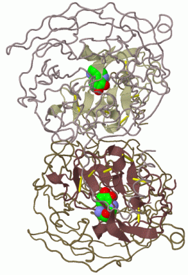
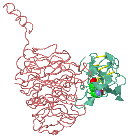
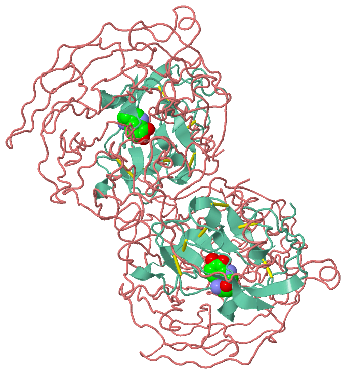
Asymmetric Unit (1, 1)
No.NameCountTypeFull Name
1TRQ1Mod. Amino Acid2-AMINO-3-(6,7-DIOXO-6,7-DIHYDRO-1H-INDOL-3-YL)-PROPIONIC ACID
Biological Unit 1 (1, 2)
No.NameCountTypeFull Name
1TRQ2Mod. Amino Acid2-AMINO-3-(6,7-DIOXO-6,7-DIHYDRO-1H-INDOL-3-YL)-PROPIONIC ACID

(-) Sites  (0, 0)

(no "Site" information available for 2MAD)

(-) SS Bonds  (6, 6)

Asymmetric Unit
No.Residues
1L:23 -L:88
2L:29 -L:61
3L:36 -L:121
4L:38 -L:86
5L:46 -L:77
6L:78 -L:109

(-) Cis Peptide Bonds  (1, 1)

Asymmetric Unit
No.Residues
1Lys L:129 -Ala L:130

 Sequence-Structure Mapping

(-) SAPs(SNPs)/Variants  (0, 0)

(no "SAP(SNP)/Variant" information available for 2MAD)

(-) PROSITE Motifs  (0, 0)

(no "PROSITE Motif" information available for 2MAD)

(-) Exons   (0, 0)

(no "Exon" information available for 2MAD)

(-) Sequences/Alignments

Asymmetric Unit
   Reformat: Number of residues per line =  ('0' or empty: single-line sequence representation)
  Number of residues per labelling interval =   
  UniProt sequence: complete  aligned part    
   Show mapping: SCOP domains CATH domains Pfam domains Secondary structure (by author)
SAPs(SNPs) PROSITE motifs Exons
(details for a mapped element are shown in a popup box when the mouse pointer rests over it)
Chain H from PDB  Type:PROTEIN  Length:373
                                                                                                                                                                                                                                                                                                                                                                                                                     
               SCOP domains d2madh_ H: Methylamine dehydrogenase, H-chain                                                                                                                                                                                                                                                                                                                                         SCOP domains
               CATH domains ------------------------------------------------------------------------------------------------------------------------------------------------------------------------------------------------------------------------------------------------------------------------------------------------------------------------------------------------------------------------------------- CATH domains
               Pfam domains ------------------------------------------------------------------------------------------------------------------------------------------------------------------------------------------------------------------------------------------------------------------------------------------------------------------------------------------------------------------------------------- Pfam domains
         Sec.struct. author ..................................................................................................................................................................................................................................................................................................................................................................................... Sec.struct. author
                 SAPs(SNPs) ------------------------------------------------------------------------------------------------------------------------------------------------------------------------------------------------------------------------------------------------------------------------------------------------------------------------------------------------------------------------------------- SAPs(SNPs)
                    PROSITE ------------------------------------------------------------------------------------------------------------------------------------------------------------------------------------------------------------------------------------------------------------------------------------------------------------------------------------------------------------------------------------- PROSITE
                 Transcript ------------------------------------------------------------------------------------------------------------------------------------------------------------------------------------------------------------------------------------------------------------------------------------------------------------------------------------------------------------------------------------- Transcript
                 2mad H   1 SSASAAAAAAAAALAAGAADGPTNDEAPGADGRRSYINLPAHHSAIIQQWVLDAGSGSILGHVNGGFLPNPVAAHSGSEFALASTSFSRIAKGKRTDYVEVFDPVTFLPIADIELPDAPRFDVGPYSWMNANTPNNADLLFFQFAAGPAVGLVVQGGSSDDQLLSSPTCYHIHPGAPSTFYLLCAQGGLAKTDHAGGAAGAGLVGAMLTAAQNLLTQPAQANKSGRIVWPVYSGKILQADISAAGATNKAPIDALSGGRKADTWRPGGWQQVAYLKSSDGIYLLTSEQSAWKLHAAAKEVTSVTGLVGQTSSQISLGHDVDAISVAQDGGPDLYALSAGTEVLHIYDAGAGDQDQSTVELGSGPQVLSVMNEA 373
                                    10        20        30        40        50        60        70        80        90       100       110       120       130       140       150       160       170       180       190       200       210       220       230       240       250       260       270       280       290       300       310       320       330       340       350       360       370   

Chain L from PDB  Type:PROTEIN  Length:124
 aligned with DHML_PARVE | P22641 from UniProtKB/Swiss-Prot  Length:188

    Alignment length:124
                                    73        83        93       103       113       123       133       143       153       163       173       183    
           DHML_PARVE    64 VDPRAKWQPQDNDIQACDYWRHCSIDGNICDCSGGSLTNCPPGTKLATASWVASCYNPTDGQSYLIAYRDCCGYNVSGRCPCLNTEGELPVYRPEFANDIIWCFGAEDDAMTYHCTISPIVGKA 187
               SCOP domains d2madl_ L: Methylamine dehydrogenase                                                                                         SCOP domains
               CATH domains 2madL00 L:7-130 Electron Transport Ethylamine Dehydrogenase                                                                  CATH domains
               Pfam domains -Me-amine-dh_L-2madL01 L:8-130                                                                                               Pfam domains
         Sec.struct. author ...................hhhh..eee.hhhh...........ee...............eeeeeee.eee........eee.........hhhhh........hhhhh..eeee...eeee. Sec.struct. author
                 SAPs(SNPs) ---------------------------------------------------------------------------------------------------------------------------- SAPs(SNPs)
                    PROSITE ---------------------------------------------------------------------------------------------------------------------------- PROSITE
                 Transcript ---------------------------------------------------------------------------------------------------------------------------- Transcript
                 2mad L   7 VDPRAKWQPQDNDIQACDYWRHCSIDGNICDCSGGSLTNCPPGTKLATASwVASCYNPTDGQSYLIAYRDCCGYNVSGRCPCLNTEGELPVYRPEFANDIIWCFGAEDDAMTYHCTISPIVGKA 130
                                    16        26        36        46        56|       66        76        86        96       106       116       126    
                                                                             57-TRQ                                                                     

   Legend:   → Mismatch (orange background)
  - → Gap (green background, '-', border residues have a numbering label)
    → Modified Residue (blue background, lower-case, 'x' indicates undefined single-letter code, labelled with number + name)
  x → Chemical Group (purple background, 'x', labelled with number + name, e.g. ACE or NH2)
  extra numbering lines below/above indicate numbering irregularities and modified residue names etc., number ends below/above '|'

 Classification and Annotation

(-) SCOP Domains  (2, 2)

Asymmetric Unit

(-) CATH Domains  (1, 1)

Asymmetric Unit
(-)
Class: Mainly Beta (13760)

(-) Pfam Domains  (1, 1)

Asymmetric Unit

(-) Gene Ontology  (9, 14)

Asymmetric Unit(hide GO term definitions)
Chain L   (DHML_PARVE | P22641)
molecular function
    GO:0030058    amine dehydrogenase activity    Catalysis of the reaction: R-CH2-NH2 + H2O + acceptor = R-CHO + NH3 + reduced acceptor.
    GO:0052876    methylamine dehydrogenase (amicyanin) activity    Catalysis of the reaction: methylamine + H2O + amicyanin = formaldehyde + ammonia + reduced amicyanin.
    GO:0016491    oxidoreductase activity    Catalysis of an oxidation-reduction (redox) reaction, a reversible chemical reaction in which the oxidation state of an atom or atoms within a molecule is altered. One substrate acts as a hydrogen or electron donor and becomes oxidized, while the other acts as hydrogen or electron acceptor and becomes reduced.
    GO:0016638    oxidoreductase activity, acting on the CH-NH2 group of donors    Catalysis of an oxidation-reduction (redox) reaction in which a CH-NH2 group acts as a hydrogen or electron donor and reduces a hydrogen or electron acceptor.
biological process
    GO:0009308    amine metabolic process    The chemical reactions and pathways involving any organic compound that is weakly basic in character and contains an amino or a substituted amino group. Amines are called primary, secondary, or tertiary according to whether one, two, or three carbon atoms are attached to the nitrogen atom.
    GO:0055114    oxidation-reduction process    A metabolic process that results in the removal or addition of one or more electrons to or from a substance, with or without the concomitant removal or addition of a proton or protons.
cellular component
    GO:0030288    outer membrane-bounded periplasmic space    The region between the inner (cytoplasmic or plasma) membrane and outer membrane of organisms with two membranes such as Gram negative bacteria. These periplasmic spaces are relatively thick and contain a thin peptidoglycan layer (PGL), also referred to as a thin cell wall.
    GO:0042597    periplasmic space    The region between the inner (cytoplasmic) and outer membrane (Gram-negative Bacteria) or cytoplasmic membrane and cell wall (Fungi and Gram-positive Bacteria).

 Visualization

(-) Interactive Views

Asymmetric Unit
  Complete Structure
    Jena3D(integrated viewing of ligand, site, SAP, PROSITE, SCOP information)
    WebMol | AstexViewer[tm]@PDBe
(Java Applets, require no local installation except for Java; loading may be slow)
    STRAP
(Java WebStart application, automatic local installation, requires Java; full application with system access!)
    RasMol
(require local installation)
    Molscript (VRML)
(requires installation of a VRML viewer; select preferred view via VRML and generate a mono or stereo PDF format file)
 
  Ligands, Modified Residues, Ions
    TRQ  [ RasMol | Jena3D ]  +environment [ RasMol | Jena3D ]
 
  Sites
(no "Sites" information available for 2mad)
 
  Cis Peptide Bonds
    Lys L:129 - Ala L:130   [ RasMol ]  
 
Biological Unit
  Complete Structure
    Biological Unit 1  [ Jena3D ]

(-) Still Images

Jmol
  protein: cartoon or spacefill or dots and stick; nucleic acid: cartoon and stick; ligands: spacefill; active site: stick
Molscript
  protein, nucleic acid: cartoon; ligands: spacefill; active site: ball and stick

 Databases and Analysis Tools

(-) Databases

Access by PDB/NDB ID
  2mad
    Family and Domain InformationProDom | SYSTERS
    General Structural InformationGlycoscienceDB | MMDB | NDB | OCA | PDB | PDBe | PDBj | PDBsum | PDBWiki | PQS | PROTEOPEDIA
    Orientation in MembranesOPM
    Protein SurfaceSURFACE
    Secondary StructureDSSP (structure derived) | HSSP (homology derived)
    Structural GenomicsGeneCensus
    Structural NeighboursCE | VAST
    Structure ClassificationCATH | Dali | SCOP
    Validation and Original DataBMRB Data View | BMRB Restraints Grid | EDS | PROCHECK | RECOORD | WHAT_CHECK
 
Access by UniProt ID/Accession number
  DHMH_PARVE | P23006
    Comparative Protein Structure ModelsModBase
    Genomic InformationEnsembl
    Protein-protein InteractionDIP
    Sequence, Family and Domain InformationInterPro | Pfam | SMART | UniProtKB/SwissProt
  DHML_PARVE | P22641
    Comparative Protein Structure ModelsModBase
    Genomic InformationEnsembl
    Protein-protein InteractionDIP
    Sequence, Family and Domain InformationInterPro | Pfam | SMART | UniProtKB/SwissProt
 
Access by Enzyme Classificator   (EC Number)
  1.4.99.3
    General Enzyme InformationBRENDA | EC-PDB | Enzyme | IntEnz
    PathwayKEGG | MetaCyc
 
Access by Disease Identifier   (MIM ID)
  (no 'MIM ID' available)
    Disease InformationOMIM
 
Access by GenAge ID
  (no 'GenAge ID' available)
    Age Related InformationGenAge

(-) Analysis Tools

Access by PDB/NDB ID
    Domain InformationXDom
    Interatomic Contacts of Structural UnitsCSU
    Ligand-protein ContactsLPC
    Protein CavitiescastP
    Sequence and Secondary StructurePDBCartoon
    Structure AlignmentSTRAP(Java WebStart application, automatic local installation, requires Java; full application with system access!)
    Structure and Sequence BrowserSTING
 
Access by UniProt ID/Accession number
  DHMH_PARVE | P23006
    Protein Disorder PredictionDisEMBL | FoldIndex | GLOBPLOT (for more information see DisProt)
  DHML_PARVE | P22641
    Protein Disorder PredictionDisEMBL | FoldIndex | GLOBPLOT (for more information see DisProt)

 Related Entries

(-) Entries Sharing at Least One Protein Chain (UniProt ID)

UniProtKB/Swiss-Prot
        DHMH_PARVE | P230061mae 1maf 3c75
        DHML_PARVE | P226411mae 1maf 3c75

(-) Related Entries Specified in the PDB File

(no "Related Entries Specified in the PDB File" available for 2MAD)