| molecular function |
|---|
| | GO:0008289 | | lipid binding | | Interacting selectively and non-covalently with a lipid. |
| | GO:0005547 | | phosphatidylinositol-3,4,5-trisphosphate binding | | Interacting selectively and non-covalently with phosphatidylinositol-3,4,5-trisphosphate, a derivative of phosphatidylinositol in which the inositol ring is phosphorylated at the 3', 4' and 5' positions. |
| | GO:0005515 | | protein binding | | Interacting selectively and non-covalently with any protein or protein complex (a complex of two or more proteins that may include other nonprotein molecules). |
| biological process |
|---|
| | GO:0016055 | | Wnt signaling pathway | | The series of molecular signals initiated by binding of a Wnt protein to a frizzled family receptor on the surface of the target cell and ending with a change in cell state. |
| | GO:0007155 | | cell adhesion | | The attachment of a cell, either to another cell or to an underlying substrate such as the extracellular matrix, via cell adhesion molecules. |
| | GO:0034329 | | cell junction assembly | | A cellular process that results in the aggregation, arrangement and bonding together of a set of components to form a cell junction. |
| | GO:0007160 | | cell-matrix adhesion | | The binding of a cell to the extracellular matrix via adhesion molecules. |
| | GO:0048041 | | focal adhesion assembly | | The aggregation and bonding together of a set of components to form a focal adhesion, a complex of intracellular signaling and structural proteins that provides a structural link between the internal actin cytoskeleton and the ECM, and also function as a locus of signal transduction activity. |
| | GO:0033622 | | integrin activation | | The aggregation, arrangement and bonding together of an integrin, a heterodimeric adhesion receptor formed by the non-covalent association of particular alpha and beta subunits, that lead to the increased affinity of the integrin for its extracellular ligands. |
| | GO:0007229 | | integrin-mediated signaling pathway | | A series of molecular signals initiated by the binding of extracellular ligand to an integrin on the surface of a target cell, and ending with regulation of a downstream cellular process, e.g. transcription. |
| | GO:0072657 | | protein localization to membrane | | A process in which a protein is transported to, or maintained in, a specific location in a membrane. |
| | GO:0008360 | | regulation of cell shape | | Any process that modulates the surface configuration of a cell. |
| | GO:0034446 | | substrate adhesion-dependent cell spreading | | The morphogenetic process that results in flattening of a cell as a consequence of its adhesion to a substrate. |
| | GO:0007179 | | transforming growth factor beta receptor signaling pathway | | A series of molecular signals initiated by the binding of an extracellular ligand to a transforming growth factor beta receptor on the surface of a target cell, and ending with regulation of a downstream cellular process, e.g. transcription. |
| cellular component |
|---|
| | GO:0031674 | | I band | | A region of a sarcomere that appears as a light band on each side of the Z disc, comprising a region of the sarcomere where thin (actin) filaments are not overlapped by thick (myosin) filaments; contains actin, troponin, and tropomyosin; each sarcomere includes half of an I band at each end. |
| | GO:0005938 | | cell cortex | | The region of a cell that lies just beneath the plasma membrane and often, but not always, contains a network of actin filaments and associated proteins. |
| | GO:0030054 | | cell junction | | A cellular component that forms a specialized region of connection between two or more cells or between a cell and the extracellular matrix. At a cell junction, anchoring proteins extend through the plasma membrane to link cytoskeletal proteins in one cell to cytoskeletal proteins in neighboring cells or to proteins in the extracellular matrix. |
| | GO:0042995 | | cell projection | | A prolongation or process extending from a cell, e.g. a flagellum or axon. |
| | GO:0009986 | | cell surface | | The external part of the cell wall and/or plasma membrane. |
| | GO:0005737 | | cytoplasm | | All of the contents of a cell excluding the plasma membrane and nucleus, but including other subcellular structures. |
| | GO:0005856 | | cytoskeleton | | Any of the various filamentous elements that form the internal framework of cells, and typically remain after treatment of the cells with mild detergent to remove membrane constituents and soluble components of the cytoplasm. The term embraces intermediate filaments, microfilaments, microtubules, the microtrabecular lattice, and other structures characterized by a polymeric filamentous nature and long-range order within the cell. The various elements of the cytoskeleton not only serve in the maintenance of cellular shape but also have roles in other cellular functions, including cellular movement, cell division, endocytosis, and movement of organelles. |
| | GO:0005829 | | cytosol | | The part of the cytoplasm that does not contain organelles but which does contain other particulate matter, such as protein complexes. |
| | GO:0031234 | | extrinsic component of cytoplasmic side of plasma membrane | | The component of a plasma membrane consisting of gene products and protein complexes that are loosely bound to its cytoplasmic surface, but not integrated into the hydrophobic region. |
| | GO:0031941 | | filamentous actin | | A two-stranded helical polymer of the protein actin. |
| | GO:0005925 | | focal adhesion | | Small region on the surface of a cell that anchors the cell to the extracellular matrix and that forms a point of termination of actin filaments. |
| | GO:0031258 | | lamellipodium membrane | | The portion of the plasma membrane surrounding a lamellipodium. |
| | GO:0016020 | | membrane | | A lipid bilayer along with all the proteins and protein complexes embedded in it an attached to it. |
| | GO:0005654 | | nucleoplasm | | That part of the nuclear content other than the chromosomes or the nucleolus. |
| | GO:0005634 | | nucleus | | A membrane-bounded organelle of eukaryotic cells in which chromosomes are housed and replicated. In most cells, the nucleus contains all of the cell's chromosomes except the organellar chromosomes, and is the site of RNA synthesis and processing. In some species, or in specialized cell types, RNA metabolism or DNA replication may be absent. |
| | GO:0005886 | | plasma membrane | | The membrane surrounding a cell that separates the cell from its external environment. It consists of a phospholipid bilayer and associated proteins. |
| | GO:0001725 | | stress fiber | | A contractile actin filament bundle that consists of short actin filaments with alternating polarity, cross-linked by alpha-actinin and possibly other actin bundling proteins, and with myosin present in a periodic distribution along the fiber. |
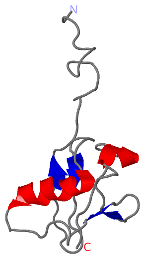
 Description
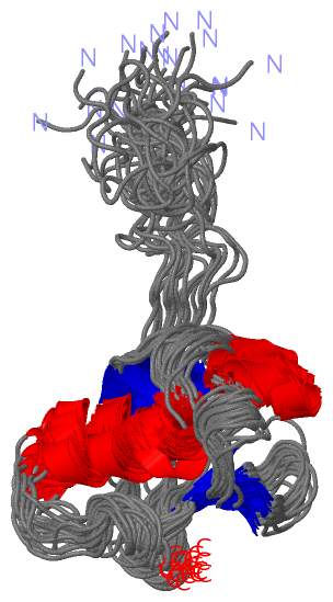