Show PDB file:   
         Plain Text   HTML   (compressed file size)
QuickSearch:   
by PDB,NDB,UniProt,PROSITE Code or Search Term(s)  
(-)NMR Structure - model 1
(-)NMR Structure - model 1, sites
(-)NMR Structure - all models
collapse expand < >
Image NMR Structure - model 1
NMR Structure - model 1  (Jmol Viewer)
Image NMR Structure - model 1, sites
NMR Structure - model 1, sites  (Jmol Viewer)
Image NMR Structure - all models
NMR Structure - all models  (Jmol Viewer)

(-) Description

Title :  REDOX LINKED CONFORMATIONAL CHANGES IN CYTOCHROME C3 FROM DESULFOVIBRIO DESULFURICANS ATCC 27774
 
Authors :  D. L. Turner, V. B. Paixao
Date :  13 Jan 10  (Deposition) - 15 Sep 10  (Release) - 14 Aug 13  (Revision)
Method :  SOLUTION NMR
Resolution :  NOT APPLICABLE
Chains :  NMR Structure  :  A  (20x)
NMR Structure *:  A  (1x)
Keywords :  Desulfovibrio Desulfuricans, Atcc 27774, Multihaem Cytochrome, Fully Reduced, Electron Transport (Keyword Search: [Gene Ontology, PubMed, Web (Google))
 
Reference :  V. B. Paixao, H. Vis, D. L. Turner
Redox Linked Conformational Changes In Cytochrome C3 From Desulfovibrio Desulfuricans Atcc 27774
Biochemistry V. 49 9620 2010
PubMed-ID: 20886839  |  Reference-DOI: 10.1021/BI101237W

(-) Compounds

Molecule 1 - CYTOCHROME C3
    ChainsA
    Organism ScientificDESULFOVIBRIO DESULFURICANS
    Organism Taxid525146
    Other DetailsFULLY REDUCED PROTEIN
    StrainATCC 27774

 Structural Features

(-) Chains, Units

  1
NMR Structure (20x)A
NMR Structure * (1x)A

Summary Information (see also Sequences/Alignments below)

(-) Ligands, Modified Residues, Ions  (1, 4)

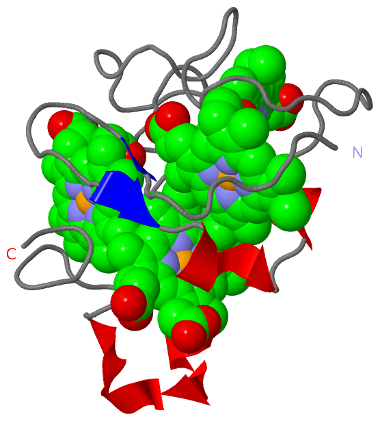
NMR Structure (1, 4)
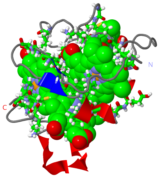
No.NameCountTypeFull Name
1HEM4Ligand/IonPROTOPORPHYRIN IX CONTAINING FE
NMR Structure * (1, 4)
No.NameCountTypeFull Name
1HEM4Ligand/IonPROTOPORPHYRIN IX CONTAINING FE

(-) Sites  (4, 4)

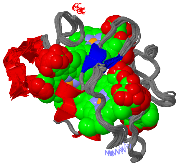
NMR Structure (4, 4)
No.NameEvidenceResiduesDescription
1AC1SOFTWAREPRO A:2 , VAL A:11 , PHE A:20 , HIS A:22 , HIS A:25 , VAL A:28 , GLU A:29 , CYS A:30 , CYS A:33 , HIS A:34 , TYR A:43 , ALA A:44 , LYS A:45 , CYS A:46 , HEM A:282BINDING SITE FOR RESIDUE HEM A 233
2AC2SOFTWAREHIS A:35 , ALA A:44 , LYS A:45 , CYS A:46 , GLY A:50 , CYS A:51 , HIS A:52 , TYR A:66 , VAL A:67 , LEU A:74 , LYS A:75BINDING SITE FOR RESIDUE HEM A 251
3AC3SOFTWAREPHE A:20 , PRO A:24 , HIS A:25 , VAL A:28 , CYS A:79 , CYS A:82 , HIS A:83 , LYS A:104 , HEM A:233BINDING SITE FOR RESIDUE HEM A 282
4AC4SOFTWAREVAL A:11 , GLY A:13 , SER A:14 , LEU A:55 , LEU A:64 , TYR A:65 , VAL A:68 , HIS A:69 , HIS A:83 , LEU A:97 , GLY A:99 , CYS A:100 , CYS A:105 , HIS A:106BINDING SITE FOR RESIDUE HEM A 305

(-) SS Bonds  (0, 0)

(no "SS Bond" information available for 2KSU)

(-) Cis Peptide Bonds  (0, 0)

(no "Cis Peptide Bond" information available for 2KSU)

 Sequence-Structure Mapping

(-) SAPs(SNPs)/Variants  (0, 0)

(no "SAP(SNP)/Variant" information available for 2KSU)

(-) PROSITE Motifs  (0, 0)

(no "PROSITE Motif" information available for 2KSU)

(-) Exons   (0, 0)

(no "Exon" information available for 2KSU)

(-) Sequences/Alignments

NMR Structure
   Reformat: Number of residues per line =  ('0' or empty: single-line sequence representation)
  Number of residues per labelling interval =   
  UniProt sequence: complete  aligned part    
   Show mapping: SCOP domains CATH domains Pfam domains Secondary structure (by author)
SAPs(SNPs) PROSITE motifs Exons
(details for a mapped element are shown in a popup box when the mouse pointer rests over it)
Chain A from PDB  Type:PROTEIN  Length:107
 aligned with B8J2Z0_DESDA | B8J2Z0 from UniProtKB/TrEMBL  Length:128

    Alignment length:107
                                    31        41        51        61        71        81        91       101       111       121       
         B8J2Z0_DESDA    22 APAVPDKPVEVKGSQKTVMFPHAPHEKVECVTCHHLVDGKESYAKCGSSGCHDDLTAKKGEKSLYYVVHAKGELKHTSCLACHSKVVAEKPELKKDLTGCAKSKCHP 128
               SCOP domains d2ksua_ A: Cytochrome c3                                                                                    SCOP domains
               CATH domains 2ksuA00 A:1-107 Cytochrome C3                                                                               CATH domains
               Pfam domains Cytochrom_CIII-2ksuA01 A:1-106                                                                            - Pfam domains
         Sec.struct. author ........ee........ee.hhhhh..hhhhhh...........................................hhhhhhhhhhhhhhhhh............. Sec.struct. author
                 SAPs(SNPs) ----------------------------------------------------------------------------------------------------------- SAPs(SNPs)
                    PROSITE ----------------------------------------------------------------------------------------------------------- PROSITE
                 Transcript ----------------------------------------------------------------------------------------------------------- Transcript
                 2ksu A   1 APAVPDKPVEVKGSQKTVMFPHAPHEKVECVTCHHLVDGKESYAKCGSSGCHDDLTAKKGEKSLYYVVHARGELKHTSCLACHSKVVAEKPELKKDLTGCAKSKCHP 107
                                    10        20        30        40        50        60        70        80        90       100       

Chain A from PDB  Type:PROTEIN  Length:107
 aligned with Q9L915_DESDE | Q9L915 from UniProtKB/TrEMBL  Length:128

    Alignment length:107
                                    31        41        51        61        71        81        91       101       111       121       
         Q9L915_DESDE    22 APAVPDKPVEVKGSQKTVMFPHAPHEKVECVTCHHLVDGKESYAKCGSSGCHDDLTAKKGEKSLYYVVHAKGELKHTSCLACHSKVVAEKPELKKDLTGCAKSKCHP 128
               SCOP domains d2ksua_ A: Cytochrome c3                                                                                    SCOP domains
               CATH domains 2ksuA00 A:1-107 Cytochrome C3                                                                               CATH domains
               Pfam domains Cytochrom_CIII-2ksuA01 A:1-106                                                                            - Pfam domains
         Sec.struct. author ........ee........ee.hhhhh..hhhhhh...........................................hhhhhhhhhhhhhhhhh............. Sec.struct. author
                 SAPs(SNPs) ----------------------------------------------------------------------------------------------------------- SAPs(SNPs)
                    PROSITE ----------------------------------------------------------------------------------------------------------- PROSITE
                 Transcript ----------------------------------------------------------------------------------------------------------- Transcript
                 2ksu A   1 APAVPDKPVEVKGSQKTVMFPHAPHEKVECVTCHHLVDGKESYAKCGSSGCHDDLTAKKGEKSLYYVVHARGELKHTSCLACHSKVVAEKPELKKDLTGCAKSKCHP 107
                                    10        20        30        40        50        60        70        80        90       100       

   Legend:   → Mismatch (orange background)
  - → Gap (green background, '-', border residues have a numbering label)
    → Modified Residue (blue background, lower-case, 'x' indicates undefined single-letter code, labelled with number + name)
  x → Chemical Group (purple background, 'x', labelled with number + name, e.g. ACE or NH2)
  extra numbering lines below/above indicate numbering irregularities and modified residue names etc., number ends below/above '|'

 Classification and Annotation

(-) SCOP Domains  (1, 1)

NMR Structure

(-) CATH Domains  (1, 1)

NMR Structure
(-)
Class: Alpha Beta (26913)

(-) Pfam Domains  (1, 1)

NMR Structure

(-) Gene Ontology  (3, 6)

NMR Structure(hide GO term definitions)
Chain A   (Q9L915_DESDE | Q9L915)
molecular function
    GO:0009055    electron carrier activity    Any molecular entity that serves as an electron acceptor and electron donor in an electron transport chain. An electron transport chain is a process in which a series of electron carriers operate together to transfer electrons from donors to any of several different terminal electron acceptors to generate a transmembrane electrochemical gradient.
    GO:0020037    heme binding    Interacting selectively and non-covalently with heme, any compound of iron complexed in a porphyrin (tetrapyrrole) ring.
    GO:0046872    metal ion binding    Interacting selectively and non-covalently with any metal ion.

Chain A   (B8J2Z0_DESDA | B8J2Z0)
molecular function
    GO:0009055    electron carrier activity    Any molecular entity that serves as an electron acceptor and electron donor in an electron transport chain. An electron transport chain is a process in which a series of electron carriers operate together to transfer electrons from donors to any of several different terminal electron acceptors to generate a transmembrane electrochemical gradient.
    GO:0020037    heme binding    Interacting selectively and non-covalently with heme, any compound of iron complexed in a porphyrin (tetrapyrrole) ring.
    GO:0046872    metal ion binding    Interacting selectively and non-covalently with any metal ion.

 Visualization

(-) Interactive Views

NMR Structure
  Complete Structure
    Jena3D(integrated viewing of ligand, site, SAP, PROSITE, SCOP information)
    WebMol | AstexViewer[tm]@PDBe
(Java Applets, require no local installation except for Java; loading may be slow)
    STRAP
(Java WebStart application, automatic local installation, requires Java; full application with system access!)
    RasMol
(require local installation)
    Molscript (VRML)
(requires installation of a VRML viewer; select preferred view via VRML and generate a mono or stereo PDF format file)
 
  Ligands, Modified Residues, Ions
    HEM  [ RasMol | Jena3D ]  +environment [ RasMol | Jena3D ]
 
  Sites
    AC1  [ RasMol ]  +environment [ RasMol ]
    AC2  [ RasMol ]  +environment [ RasMol ]
    AC3  [ RasMol ]  +environment [ RasMol ]
    AC4  [ RasMol ]  +environment [ RasMol ]
 
  Cis Peptide Bonds
(no "Cis Peptide Bonds" information available for 2ksu)
 

(-) Still Images

Jmol
  protein: cartoon or spacefill or dots and stick; nucleic acid: cartoon and stick; ligands: spacefill; active site: stick
Molscript
  protein, nucleic acid: cartoon; ligands: spacefill; active site: ball and stick

 Databases and Analysis Tools

(-) Databases

Access by PDB/NDB ID
  2ksu
    Family and Domain InformationProDom | SYSTERS
    General Structural InformationGlycoscienceDB | MMDB | NDB | OCA | PDB | PDBe | PDBj | PDBsum | PDBWiki | PQS | PROTEOPEDIA
    Orientation in MembranesOPM
    Protein SurfaceSURFACE
    Secondary StructureDSSP (structure derived) | HSSP (homology derived)
    Structural GenomicsGeneCensus
    Structural NeighboursCE | VAST
    Structure ClassificationCATH | Dali | SCOP
    Validation and Original DataBMRB Data View | BMRB Restraints Grid | EDS | PROCHECK | RECOORD | WHAT_CHECK
 
Access by UniProt ID/Accession number
  B8J2Z0_DESDA | B8J2Z0
    Comparative Protein Structure ModelsModBase
    Genomic InformationEnsembl
    Protein-protein InteractionDIP
    Sequence, Family and Domain InformationInterPro | Pfam | SMART | UniProtKB/TrEMBL
  Q9L915_DESDE | Q9L915
    Comparative Protein Structure ModelsModBase
    Genomic InformationEnsembl
    Protein-protein InteractionDIP
    Sequence, Family and Domain InformationInterPro | Pfam | SMART | UniProtKB/SwissProt
 
Access by Enzyme Classificator   (EC Number)
  (no 'Enzyme Classificator' available)
    General Enzyme InformationBRENDA | EC-PDB | Enzyme | IntEnz
    PathwayKEGG | MetaCyc
 
Access by Disease Identifier   (MIM ID)
  (no 'MIM ID' available)
    Disease InformationOMIM
 
Access by GenAge ID
  (no 'GenAge ID' available)
    Age Related InformationGenAge

(-) Analysis Tools

Access by PDB/NDB ID
    Domain InformationXDom
    Interatomic Contacts of Structural UnitsCSU
    Ligand-protein ContactsLPC
    Protein CavitiescastP
    Sequence and Secondary StructurePDBCartoon
    Structure AlignmentSTRAP(Java WebStart application, automatic local installation, requires Java; full application with system access!)
    Structure and Sequence BrowserSTING
 
Access by UniProt ID/Accession number
  B8J2Z0_DESDA | B8J2Z0
    Protein Disorder PredictionDisEMBL | FoldIndex | GLOBPLOT (for more information see DisProt)
  Q9L915_DESDE | Q9L915
    Protein Disorder PredictionDisEMBL | FoldIndex | GLOBPLOT (for more information see DisProt)

 Related Entries

(-) Entries Sharing at Least One Protein Chain (UniProt ID)

UniProtKB/Swiss-Prot
        Q9L915_DESDE | Q9L9152kmy
UniProtKB/TrEMBL
        B8J2Z0_DESDA | B8J2Z02kmy
        Q9L915_DESDE | Q9L9151gm4 1gmb 1i77 1up9 1upd 3cyr

(-) Related Entries Specified in the PDB File

2kmy