Show PDB file:   
         Plain Text   HTML   (compressed file size)
QuickSearch:   
by PDB,NDB,UniProt,PROSITE Code or Search Term(s)  
(-)NMR Structure - model 1
(-)NMR Structure - model 1, sites
(-)NMR Structure - all models
collapse expand < >
Image NMR Structure - model 1
NMR Structure - model 1  (Jmol Viewer)
Image NMR Structure - model 1, sites
NMR Structure - model 1, sites  (Jmol Viewer)
Image NMR Structure - all models
NMR Structure - all models  (Jmol Viewer)

(-) Description

Title :  CA-S100A1 INTERACTING WITH TRTK12
 
Authors :  N. T. Wright, K. M. Varney, B. R. Cannon, M. Morgan, D. J. Weber
Date :  02 Dec 08  (Deposition) - 10 Feb 09  (Release) - 10 Feb 09  (Revision)
Method :  SOLUTION NMR
Resolution :  NOT APPLICABLE
Chains :  NMR Structure  :  A,B,X,Y  (20x)
Keywords :  S100, Ef-Hand, Protein-Protein Interaction, Conformational Change, Capz, Acetylation, Actin Capping, Actin-Binding, Calcium, Cytoplasm, Metal-Binding, Zinc, Metal Binding Protein (Keyword Search: [Gene Ontology, PubMed, Web (Google))
 
Reference :  N. T. Wright, M. T. Morgan, B. R. Cannon, K. M. Varney, D. J. Weber
Solution Structure Of Ca-S100A1-Trtk12
To Be Published
PubMed: search
(for further references see the PDB file header)

(-) Compounds

Molecule 1 - F-ACTIN-CAPPING PROTEIN SUBUNIT ALPHA-2
    ChainsX, Y
    EngineeredYES
    Expression SystemESCHERICHIA COLI
    Expression System Taxid562
    Expression System VectorPET21A
    GeneCAPZA2
    Organism CommonRAT
    Organism ScientificRATTUS NORVEGICUS
    Organism Taxid10116
    SynonymCAPZ ALPHA-2
 
Molecule 2 - PROTEIN S100-A1
    ChainsA, B
    EngineeredYES
    Expression SystemESCHERICHIA COLI
    Expression System Taxid562
    Expression System VectorPET21A
    GeneS100A1
    Organism CommonRAT
    Organism ScientificRATTUS NORVEGICUS
    Organism Taxid10116
    SynonymS100 CALCIUM-BINDING PROTEIN A1, S-100 PROTEIN ALPHA SUBUNIT, S-100 PROTEIN ALPHA CHAIN

 Structural Features

(-) Chains, Units

  
NMR Structure (20x)

Summary Information (see also Sequences/Alignments below)

(-) Ligands, Modified Residues, Ions  (1, 4)

NMR Structure (1, 4)
No.NameCountTypeFull Name
1CA4Ligand/IonCALCIUM ION

(-) Sites  (4, 4)

NMR Structure (4, 4)
No.NameEvidenceResiduesDescription
1AC1SOFTWAREHIS A:18 , SER A:19 , GLU A:22 , GLY A:23 , ASP A:24 , LEU A:28BINDING SITE FOR RESIDUE CA A 100
2AC2SOFTWARESER B:19 , GLU B:22 , LYS B:27BINDING SITE FOR RESIDUE CA B 101
3AC3SOFTWARELYS A:27 , LEU A:61 , ASP A:62 , GLU A:63BINDING SITE FOR RESIDUE CA A 102
4AC4SOFTWAREASP B:62 , GLU B:63 , ASN B:64BINDING SITE FOR RESIDUE CA B 103

(-) SS Bonds  (0, 0)

(no "SS Bond" information available for 2KBM)

(-) Cis Peptide Bonds  (0, 0)

(no "Cis Peptide Bond" information available for 2KBM)

 Sequence-Structure Mapping

(-) SAPs(SNPs)/Variants  (0, 0)

(no "SAP(SNP)/Variant" information available for 2KBM)

(-) PROSITE Motifs  (3, 6)

NMR Structure (3, 6)
 PROSITEUniProtKBPDB
No.IDACDescriptionIDLocationCountLocation
1EF_HAND_2PS50222 EF-hand calcium-binding domain profile.S10A1_RAT50-85
 
  2A:49-84
B:49-84
2S100_CABPPS00303 S-100/ICaBP type calcium binding protein signature.S10A1_RAT58-79
 
  2A:57-78
B:57-78
3EF_HAND_1PS00018 EF-hand calcium-binding domain.S10A1_RAT63-75
 
  2A:62-74
B:62-74

(-) Exons   (2, 4)

NMR Structure (2, 4)
 ENSEMBLUniProtKBPDB
No.Transcript IDExonExon IDGenome LocationLengthIDLocationLengthCountLocationLength
1.1ENSRNOT000000170001ENSRNOE00000119000chr2:182787370-182787230141S10A1_RAT1-47472A:1-46
B:1-46
46
46
1.2ENSRNOT000000170002ENSRNOE00000119682chr2:182784961-182784818144S10A1_RAT48-94472A:47-93
B:47-93
47
47

(-) Sequences/Alignments

NMR Structure
   Reformat: Number of residues per line =  ('0' or empty: single-line sequence representation)
  Number of residues per labelling interval =   
  UniProt sequence: complete  aligned part    
   Show mapping: SCOP domains CATH domains Pfam domains Secondary structure (by author)
SAPs(SNPs) PROSITE motifs Exons
(details for a mapped element are shown in a popup box when the mouse pointer rests over it)
Chain A from PDB  Type:PROTEIN  Length:93
 aligned with S10A1_RAT | P35467 from UniProtKB/Swiss-Prot  Length:94

    Alignment length:93
                                    11        21        31        41        51        61        71        81        91   
            S10A1_RAT     2 GSELETAMETLINVFHAHSGKEGDKYKLSKKELKDLLQTELSSFLDVQKDADAVDKIMKELDENGDGEVDFQEFVVLVAALTVACNNFFWENS  94
               SCOP domains d2kbma_ A: Calcyclin (S100)                                                                   SCOP domains
               CATH domains 2kbmA00 A:1-93 EF-hand                                                                        CATH domains
               Pfam domains --------------------------------------------------------------------------------------------- Pfam domains
         Sec.struct. author .hhhhhhhhhhhhhhhhhhh.......eehhhhhhhhhhhhh....hhhhhhhhhhhhhhhh.....eehhhhhhhhhhhhhhhhhhhhh... Sec.struct. author
                 SAPs(SNPs) --------------------------------------------------------------------------------------------- SAPs(SNPs)
                PROSITE (1) ------------------------------------------------EF_HAND_2  PDB: A:49-84             --------- PROSITE (1)
                PROSITE (2) --------------------------------------------------------S100_CABP  PDB: A:57-7--------------- PROSITE (2)
                PROSITE (3) -------------------------------------------------------------EF_HAND_1    ------------------- PROSITE (3)
               Transcript 1 Exon 1.1  PDB: A:1-46 UniProt: 1-47           Exon 1.2  PDB: A:47-93 UniProt: 48-94           Transcript 1
                 2kbm A   1 GSELETAMETLINVFHAHSGKEGDKYKLSKKELKDLLQTELSSFLDVQKDADAVDKIMKELDENGDGEVDFQEFVVLVAALTVACNNFFWENS  93
                                    10        20        30        40        50        60        70        80        90   

Chain B from PDB  Type:PROTEIN  Length:93
 aligned with S10A1_RAT | P35467 from UniProtKB/Swiss-Prot  Length:94

    Alignment length:93
                                    11        21        31        41        51        61        71        81        91   
            S10A1_RAT     2 GSELETAMETLINVFHAHSGKEGDKYKLSKKELKDLLQTELSSFLDVQKDADAVDKIMKELDENGDGEVDFQEFVVLVAALTVACNNFFWENS  94
               SCOP domains d2kbmb_ B: Calcyclin (S100)                                                                   SCOP domains
               CATH domains 2kbmB00 B:1-93 EF-hand                                                                        CATH domains
           Pfam domains (1) ---S_100-2kbmB01 B:4-47                        -----efhand-2kbmB03 B:53-81       ------------ Pfam domains (1)
           Pfam domains (2) ---S_100-2kbmB02 B:4-47                        -----efhand-2kbmB04 B:53-81       ------------ Pfam domains (2)
         Sec.struct. author .hhhhhhhhhhhhhhhhhhh.......eehhhhhhhhhhhhh....hhhhhhhhhhhhhhhh.....eehhhhhhhhhhhhhhhhhhhhh... Sec.struct. author
                 SAPs(SNPs) --------------------------------------------------------------------------------------------- SAPs(SNPs)
                PROSITE (1) ------------------------------------------------EF_HAND_2  PDB: B:49-84             --------- PROSITE (1)
                PROSITE (2) --------------------------------------------------------S100_CABP  PDB: B:57-7--------------- PROSITE (2)
                PROSITE (3) -------------------------------------------------------------EF_HAND_1    ------------------- PROSITE (3)
               Transcript 1 Exon 1.1  PDB: B:1-46 UniProt: 1-47           Exon 1.2  PDB: B:47-93 UniProt: 48-94           Transcript 1
                 2kbm B   1 GSELETAMETLINVFHAHSGKEGDKYKLSKKELKDLLQTELSSFLDVQKDADAVDKIMKELDENGDGEVDFQEFVVLVAALTVACNNFFWENS  93
                                    10        20        30        40        50        60        70        80        90   

Chain X from PDB  Type:PROTEIN  Length:12
 aligned with CAZA2_RAT | Q3T1K5 from UniProtKB/Swiss-Prot  Length:286

    Alignment length:12
                                   274  
            CAZA2_RAT   265 TRTKIDWNKILS 276
               SCOP domains ------------ SCOP domains
               CATH domains ------------ CATH domains
               Pfam domains ------------ Pfam domains
         Sec.struct. author .....hhhhhhh Sec.struct. author
                 SAPs(SNPs) ------------ SAPs(SNPs)
                    PROSITE ------------ PROSITE
                 Transcript ------------ Transcript
                 2kbm X   1 TRTKIDWNKILS  12
                                    10  

Chain Y from PDB  Type:PROTEIN  Length:12
 aligned with CAZA2_RAT | Q3T1K5 from UniProtKB/Swiss-Prot  Length:286

    Alignment length:12
                                   274  
            CAZA2_RAT   265 TRTKIDWNKILS 276
               SCOP domains ------------ SCOP domains
               CATH domains ------------ CATH domains
           Pfam domains (1) F-actin_cap_ Pfam domains (1)
           Pfam domains (2) F-actin_cap_ Pfam domains (2)
         Sec.struct. author .....hhhhhhh Sec.struct. author
                 SAPs(SNPs) ------------ SAPs(SNPs)
                    PROSITE ------------ PROSITE
                 Transcript ------------ Transcript
                 2kbm Y   1 TRTKIDWNKILS  12
                                    10  

   Legend:   → Mismatch (orange background)
  - → Gap (green background, '-', border residues have a numbering label)
    → Modified Residue (blue background, lower-case, 'x' indicates undefined single-letter code, labelled with number + name)
  x → Chemical Group (purple background, 'x', labelled with number + name, e.g. ACE or NH2)
  extra numbering lines below/above indicate numbering irregularities and modified residue names etc., number ends below/above '|'

 Classification and Annotation

(-) SCOP Domains  (1, 2)

NMR Structure

(-) CATH Domains  (1, 2)

NMR Structure

(-) Pfam Domains  (3, 6)

NMR Structure
(-)
Clan: EF_hand (270)

(-) Gene Ontology  (29, 29)

NMR Structure(hide GO term definitions)
Chain A,B   (S10A1_RAT | P35467)
molecular function
    GO:0051117    ATPase binding    Interacting selectively and non-covalently with an ATPase, any enzyme that catalyzes the hydrolysis of ATP.
    GO:0044548    S100 protein binding    Interacting selectively and non-covalently with a S100 protein. S100 is a small calcium and zinc binding protein produced in astrocytes that is implicated in Alzheimer's disease, Down Syndrome and ALS.
    GO:0005509    calcium ion binding    Interacting selectively and non-covalently with calcium ions (Ca2+).
    GO:0048306    calcium-dependent protein binding    Interacting selectively and non-covalently with any protein or protein complex (a complex of two or more proteins that may include other nonprotein molecules), in the presence of calcium.
    GO:0042802    identical protein binding    Interacting selectively and non-covalently with an identical protein or proteins.
    GO:0046872    metal ion binding    Interacting selectively and non-covalently with any metal ion.
    GO:0005515    protein binding    Interacting selectively and non-covalently with any protein or protein complex (a complex of two or more proteins that may include other nonprotein molecules).
    GO:0042803    protein homodimerization activity    Interacting selectively and non-covalently with an identical protein to form a homodimer.
biological process
    GO:0000122    negative regulation of transcription from RNA polymerase II promoter    Any process that stops, prevents, or reduces the frequency, rate or extent of transcription from an RNA polymerase II promoter.
    GO:1901387    positive regulation of voltage-gated calcium channel activity    Any process that activates or increases the frequency, rate or extent of voltage-gated calcium channel activity.
    GO:0008016    regulation of heart contraction    Any process that modulates the frequency, rate or extent of heart contraction. Heart contraction is the process in which the heart decreases in volume in a characteristic way to propel blood through the body.
    GO:0021762    substantia nigra development    The progression of the substantia nigra over time from its initial formation until its mature state. The substantia nigra is the layer of gray substance that separates the posterior parts of the cerebral peduncles (tegmentum mesencephali) from the anterior parts; it normally includes a posterior compact part with many pigmented cells (pars compacta) and an anterior reticular part whose cells contain little pigment (pars reticularis).
cellular component
    GO:0031672    A band    The dark-staining region of a sarcomere, in which myosin thick filaments are present; the center is traversed by the paler H zone, which in turn contains the M line.
    GO:0031674    I band    A region of a sarcomere that appears as a light band on each side of the Z disc, comprising a region of the sarcomere where thin (actin) filaments are not overlapped by thick (myosin) filaments; contains actin, troponin, and tropomyosin; each sarcomere includes half of an I band at each end.
    GO:0031430    M band    The midline of aligned thick filaments in a sarcomere; location of specific proteins that link thick filaments. Depending on muscle type the M band consists of different numbers of M lines.
    GO:0030018    Z disc    Platelike region of a muscle sarcomere to which the plus ends of actin filaments are attached.
    GO:0005737    cytoplasm    All of the contents of a cell excluding the plasma membrane and nucleus, but including other subcellular structures.
    GO:0005829    cytosol    The part of the cytoplasm that does not contain organelles but which does contain other particulate matter, such as protein complexes.
    GO:0043005    neuron projection    A prolongation or process extending from a nerve cell, e.g. an axon or dendrite.
    GO:0005634    nucleus    A membrane-bounded organelle of eukaryotic cells in which chromosomes are housed and replicated. In most cells, the nucleus contains all of the cell's chromosomes except the organellar chromosomes, and is the site of RNA synthesis and processing. In some species, or in specialized cell types, RNA metabolism or DNA replication may be absent.
    GO:0016529    sarcoplasmic reticulum    A fine reticular network of membrane-limited elements that pervades the sarcoplasm of a muscle cell; continuous over large portions of the cell and with the nuclear envelope; that part of the endoplasmic reticulum specialized for calcium release, uptake and storage.
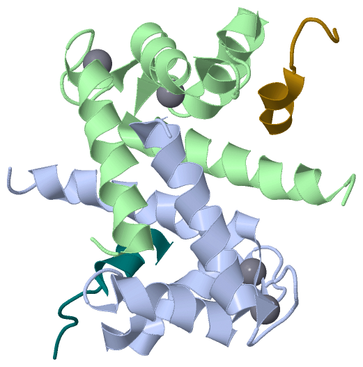
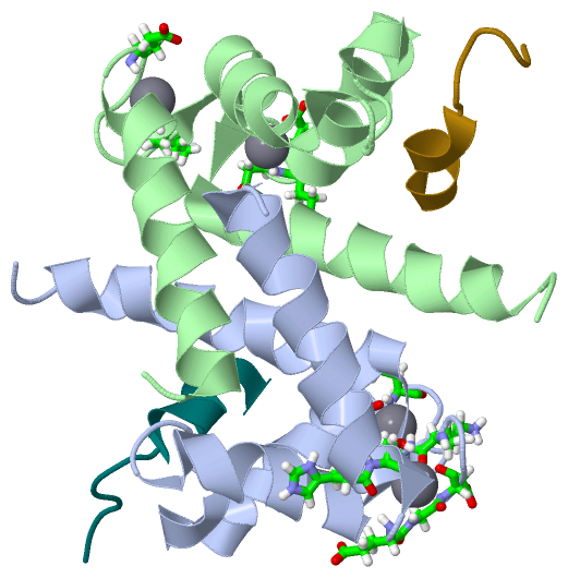
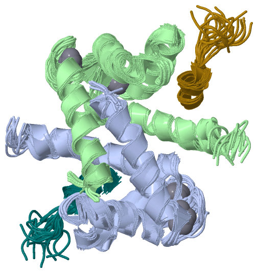
Chain X,Y   (CAZA2_RAT | Q3T1K5)
molecular function
    GO:0003779    actin binding    Interacting selectively and non-covalently with monomeric or multimeric forms of actin, including actin filaments.
biological process
    GO:0051693    actin filament capping    The binding of a protein or protein complex to the end of an actin filament, thus preventing the addition, exchange or removal of further actin subunits.
    GO:0051016    barbed-end actin filament capping    The binding of a protein or protein complex to the barbed (or plus) end of an actin filament, thus preventing the addition, exchange or removal of further actin subunits.
cellular component
    GO:0008290    F-actin capping protein complex    A heterodimer consisting of alpha and beta subunits that binds to and caps the barbed ends of actin filaments, thereby regulating the polymerization of actin monomers but not severing actin filaments.
    GO:0005903    brush border    The dense covering of microvilli on the apical surface of a epithelial cells in tissues such as the intestine, kidney, and choroid plexus; the microvilli aid absorption by increasing the surface area of the cell.
    GO:0030863    cortical cytoskeleton    The portion of the cytoskeleton that lies just beneath the plasma membrane.
    GO:0070062    extracellular exosome    A vesicle that is released into the extracellular region by fusion of the limiting endosomal membrane of a multivesicular body with the plasma membrane. Extracellular exosomes, also simply called exosomes, have a diameter of about 40-100 nm.
    GO:0016020    membrane    A lipid bilayer along with all the proteins and protein complexes embedded in it an attached to it.

 Visualization

(-) Interactive Views

NMR Structure
  Complete Structure
    Jena3D(integrated viewing of ligand, site, SAP, PROSITE, SCOP information)
    WebMol | AstexViewer[tm]@PDBe
(Java Applets, require no local installation except for Java; loading may be slow)
    STRAP
(Java WebStart application, automatic local installation, requires Java; full application with system access!)
    RasMol
(require local installation)
    Molscript (VRML)
(requires installation of a VRML viewer; select preferred view via VRML and generate a mono or stereo PDF format file)
 
  Ligands, Modified Residues, Ions
    CA  [ RasMol | Jena3D ]  +environment [ RasMol | Jena3D ]
 
  Sites
    AC1  [ RasMol ]  +environment [ RasMol ]
    AC2  [ RasMol ]  +environment [ RasMol ]
    AC3  [ RasMol ]  +environment [ RasMol ]
    AC4  [ RasMol ]  +environment [ RasMol ]
 
  Cis Peptide Bonds
(no "Cis Peptide Bonds" information available for 2kbm)
 

(-) Still Images

Jmol
  protein: cartoon or spacefill or dots and stick; nucleic acid: cartoon and stick; ligands: spacefill; active site: stick
Molscript
  protein, nucleic acid: cartoon; ligands: spacefill; active site: ball and stick

 Databases and Analysis Tools

(-) Databases

Access by PDB/NDB ID
  2kbm
    Family and Domain InformationProDom | SYSTERS
    General Structural InformationGlycoscienceDB | MMDB | NDB | OCA | PDB | PDBe | PDBj | PDBsum | PDBWiki | PQS | PROTEOPEDIA
    Orientation in MembranesOPM
    Protein SurfaceSURFACE
    Secondary StructureDSSP (structure derived) | HSSP (homology derived)
    Structural GenomicsGeneCensus
    Structural NeighboursCE | VAST
    Structure ClassificationCATH | Dali | SCOP
    Validation and Original DataBMRB Data View | BMRB Restraints Grid | EDS | PROCHECK | RECOORD | WHAT_CHECK
 
Access by UniProt ID/Accession number
  CAZA2_RAT | Q3T1K5
    Comparative Protein Structure ModelsModBase
    Genomic InformationEnsembl
    Protein-protein InteractionDIP
    Sequence, Family and Domain InformationInterPro | Pfam | SMART | UniProtKB/SwissProt
  S10A1_RAT | P35467
    Comparative Protein Structure ModelsModBase
    Genomic InformationEnsembl
    Protein-protein InteractionDIP
    Sequence, Family and Domain InformationInterPro | Pfam | SMART | UniProtKB/SwissProt
 
Access by Enzyme Classificator   (EC Number)
  (no 'Enzyme Classificator' available)
    General Enzyme InformationBRENDA | EC-PDB | Enzyme | IntEnz
    PathwayKEGG | MetaCyc
 
Access by Disease Identifier   (MIM ID)
  (no 'MIM ID' available)
    Disease InformationOMIM
 
Access by GenAge ID
  (no 'GenAge ID' available)
    Age Related InformationGenAge

(-) Analysis Tools

Access by PDB/NDB ID
    Domain InformationXDom
    Interatomic Contacts of Structural UnitsCSU
    Ligand-protein ContactsLPC
    Protein CavitiescastP
    Sequence and Secondary StructurePDBCartoon
    Structure AlignmentSTRAP(Java WebStart application, automatic local installation, requires Java; full application with system access!)
    Structure and Sequence BrowserSTING
 
Access by UniProt ID/Accession number
  CAZA2_RAT | Q3T1K5
    Protein Disorder PredictionDisEMBL | FoldIndex | GLOBPLOT (for more information see DisProt)
  S10A1_RAT | P35467
    Protein Disorder PredictionDisEMBL | FoldIndex | GLOBPLOT (for more information see DisProt)

 Related Entries

(-) Entries Sharing at Least One Protein Chain (UniProt ID)

UniProtKB/Swiss-Prot
        S10A1_RAT | P354671k2h 1zfs 2k2f

(-) Related Entries Specified in the PDB File

1zfs RELATED ID: 16050 RELATED DB: BMRB
2k2f