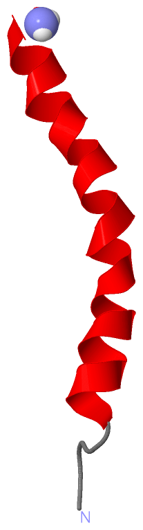
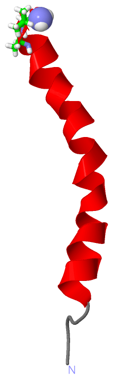
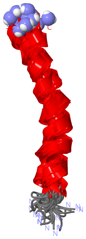
Show PDB file:   
         Plain Text   HTML   (compressed file size)
QuickSearch:   
by PDB,NDB,UniProt,PROSITE Code or Search Term(s)  
(-)NMR Structure - model 1
(-)NMR Structure - model 1, sites
(-)NMR Structure - all models
collapse expand < >
Image NMR Structure - model 1
NMR Structure - model 1  (Jmol Viewer)
Image NMR Structure - model 1, sites
NMR Structure - model 1, sites  (Jmol Viewer)
Image NMR Structure - all models
NMR Structure - all models  (Jmol Viewer)

(-) Description

Title :  STRUCTURE AND MEMBRANE INTERACTIONS OF THE ANTIBIOTIC PEPTIDE DERMADISTINCTIN K BY MULTIDIMENSIONAL SOLUTION AND ORIENTED 15N AND 31P SOLID-STATE NMR SPECTROSCOPY
 
Authors :  C. M. Moraes, R. M. Verly, J. M. Resende, C. Aisenbrey, M. P. Bemquerer, D. Pilo-Veloso, A. Valente, F. C. L. Almeida, B. Bechinger
Date :  07 Oct 08  (Deposition) - 14 Apr 09  (Release) - 14 Apr 09  (Revision)
Method :  SOLUTION NMR
Resolution :  NOT APPLICABLE
Chains :  NMR Structure  :  A  (20x)
Keywords :  Amphipathic Alpha-Helix, Membrane Protein Structure Determination, C-Terminal Carboxyamidation, Antimicrobial Protein, Amphibian Defense Peptide, Antibiotic, Antimicrobial, Secreted (Keyword Search: [Gene Ontology, PubMed, Web (Google))
 
Reference :  R. M. Verly, C. M. De Moraes, J. M. Resende, C. Aisenbrey, M. P. Bemquerer, D. Pilo-Veloso, A. P. Valente, F. C. L. Almeida, B. Bechinger
Structure And Membrane Interactions Of The Antibiotic Peptide Dermadistinctin K By Multidimensional Solution And Oriented 15N And 31P Solid-State Nmr Spectroscopy.
Biophys. J. V. 96 2194 2009
PubMed-ID: 19289046  |  Reference-DOI: 10.1016/J.BPJ.2008.11.063
(for further references see the PDB file header)

(-) Compounds

Molecule 1 - DERMADISTINCTIN-K
    ChainsA
    EngineeredYES
    Other DetailsTHE PEPTIDE WAS PREPARED BY SOLID-PHASE SYNTHESIS USING FMOC CHEMISTRY
    SynonymDD K
    SyntheticYES

 Structural Features

(-) Chains, Units

  
NMR Structure (20x)

Summary Information (see also Sequences/Alignments below)

(-) Ligands, Modified Residues, Ions  (1, 1)

NMR Structure (1, 1)
No.NameCountTypeFull Name
1NH21Mod. Amino AcidAMINO GROUP

(-) Sites  (1, 1)

NMR Structure (1, 1)
No.NameEvidenceResiduesDescription
1AC1SOFTWAREALA A:32 , VAL A:33BINDING SITE FOR RESIDUE NH2 A 34

(-) SS Bonds  (0, 0)

(no "SS Bond" information available for 2K9B)

(-) Cis Peptide Bonds  (0, 0)

(no "Cis Peptide Bond" information available for 2K9B)

 Sequence-Structure Mapping

(-) SAPs(SNPs)/Variants  (0, 0)

(no "SAP(SNP)/Variant" information available for 2K9B)

(-) PROSITE Motifs  (0, 0)

(no "PROSITE Motif" information available for 2K9B)

(-) Exons   (0, 0)

(no "Exon" information available for 2K9B)

(-) Sequences/Alignments

NMR Structure
   Reformat: Number of residues per line =  ('0' or empty: single-line sequence representation)
  Number of residues per labelling interval =   
  UniProt sequence: complete  aligned part    
   Show mapping: SCOP domains CATH domains Pfam domains Secondary structure (by author)
SAPs(SNPs) PROSITE motifs Exons
(details for a mapped element are shown in a popup box when the mouse pointer rests over it)
Chain A from PDB  Type:PROTEIN  Length:34
 aligned with DDSK_PHYDS | P83638 from UniProtKB/Swiss-Prot  Length:33

    Alignment length:34
                                                           33     
                                    10        20        30  | 
            DDSK_PHYDS    1 GLWSKIKAAGKEAAKAAAKAAGKAALNAVSEAV-  -
               SCOP domains ---------------------------------- SCOP domains
               CATH domains ---------------------------------- CATH domains
               Pfam domains ---------------------------------- Pfam domains
         Sec.struct. author ....hhhhhhhhhhhhhhhhhhhhhhhhhhhhh. Sec.struct. author
                 SAPs(SNPs) ---------------------------------- SAPs(SNPs)
                    PROSITE ---------------------------------- PROSITE
                 Transcript ---------------------------------- Transcript
                  2k9b A  1 GLWSKIKAAGKEAAKAAAKAAGKAALNAVSEAVx 34
                                    10        20        30   |
                                                            34-NH2

   Legend:   → Mismatch (orange background)
  - → Gap (green background, '-', border residues have a numbering label)
    → Modified Residue (blue background, lower-case, 'x' indicates undefined single-letter code, labelled with number + name)
  x → Chemical Group (purple background, 'x', labelled with number + name, e.g. ACE or NH2)
  extra numbering lines below/above indicate numbering irregularities and modified residue names etc., number ends below/above '|'

 Classification and Annotation

(-) SCOP Domains  (0, 0)

(no "SCOP Domain" information available for 2K9B)

(-) CATH Domains  (0, 0)

(no "CATH Domain" information available for 2K9B)

(-) Pfam Domains  (0, 0)

(no "Pfam Domain" information available for 2K9B)

(-) Gene Ontology  (3, 3)

NMR Structure(hide GO term definitions)
Chain A   (DDSK_PHYDS | P83638)
biological process
    GO:0006952    defense response    Reactions, triggered in response to the presence of a foreign body or the occurrence of an injury, which result in restriction of damage to the organism attacked or prevention/recovery from the infection caused by the attack.
    GO:0042742    defense response to bacterium    Reactions triggered in response to the presence of a bacterium that act to protect the cell or organism.
cellular component
    GO:0005576    extracellular region    The space external to the outermost structure of a cell. For cells without external protective or external encapsulating structures this refers to space outside of the plasma membrane. This term covers the host cell environment outside an intracellular parasite.

 Visualization

(-) Interactive Views

NMR Structure
  Complete Structure
    Jena3D(integrated viewing of ligand, site, SAP, PROSITE, SCOP information)
    WebMol | AstexViewer[tm]@PDBe
(Java Applets, require no local installation except for Java; loading may be slow)
    STRAP
(Java WebStart application, automatic local installation, requires Java; full application with system access!)
    RasMol
(require local installation)
    Molscript (VRML)
(requires installation of a VRML viewer; select preferred view via VRML and generate a mono or stereo PDF format file)
 
  Ligands, Modified Residues, Ions
    NH2  [ RasMol | Jena3D ]  +environment [ RasMol | Jena3D ]
 
  Sites
    AC1  [ RasMol ]  +environment [ RasMol ]
 
  Cis Peptide Bonds
(no "Cis Peptide Bonds" information available for 2k9b)
 

(-) Still Images

Jmol
  protein: cartoon or spacefill or dots and stick; nucleic acid: cartoon and stick; ligands: spacefill; active site: stick
Molscript
  protein, nucleic acid: cartoon; ligands: spacefill; active site: ball and stick

 Databases and Analysis Tools

(-) Databases

Access by PDB/NDB ID
  2k9b
    Family and Domain InformationProDom | SYSTERS
    General Structural InformationGlycoscienceDB | MMDB | NDB | OCA | PDB | PDBe | PDBj | PDBsum | PDBWiki | PQS | PROTEOPEDIA
    Orientation in MembranesOPM
    Protein SurfaceSURFACE
    Secondary StructureDSSP (structure derived) | HSSP (homology derived)
    Structural GenomicsGeneCensus
    Structural NeighboursCE | VAST
    Structure ClassificationCATH | Dali | SCOP
    Validation and Original DataBMRB Data View | BMRB Restraints Grid | EDS | PROCHECK | RECOORD | WHAT_CHECK
 
Access by UniProt ID/Accession number
  DDSK_PHYDS | P83638
    Comparative Protein Structure ModelsModBase
    Genomic InformationEnsembl
    Protein-protein InteractionDIP
    Sequence, Family and Domain InformationInterPro | Pfam | SMART | UniProtKB/SwissProt
 
Access by Enzyme Classificator   (EC Number)
  (no 'Enzyme Classificator' available)
    General Enzyme InformationBRENDA | EC-PDB | Enzyme | IntEnz
    PathwayKEGG | MetaCyc
 
Access by Disease Identifier   (MIM ID)
  (no 'MIM ID' available)
    Disease InformationOMIM
 
Access by GenAge ID
  (no 'GenAge ID' available)
    Age Related InformationGenAge

(-) Analysis Tools

Access by PDB/NDB ID
    Domain InformationXDom
    Interatomic Contacts of Structural UnitsCSU
    Ligand-protein ContactsLPC
    Protein CavitiescastP
    Sequence and Secondary StructurePDBCartoon
    Structure AlignmentSTRAP(Java WebStart application, automatic local installation, requires Java; full application with system access!)
    Structure and Sequence BrowserSTING
 
Access by UniProt ID/Accession number
  DDSK_PHYDS | P83638
    Protein Disorder PredictionDisEMBL | FoldIndex | GLOBPLOT (for more information see DisProt)

 Related Entries

(-) Entries Sharing at Least One Protein Chain (UniProt ID)

UniProtKB/Swiss-Prot
        DDSK_PHYDS | P836382jx6

(-) Related Entries Specified in the PDB File

(no "Related Entries Specified in the PDB File" available for 2K9B)