| molecular function |
|---|
| | GO:0001965 | | G-protein alpha-subunit binding | | Interacting selectively and non-covalently with a G-protein alpha subunit. The alpha subunit binds a guanine nucleotide. |
| | GO:0005092 | | GDP-dissociation inhibitor activity | | Prevents the dissociation of GDP from a GTPase, thereby preventing GTP from binding. |
| | GO:0032794 | | GTPase activating protein binding | | Interacting selectively and non-covalently with a GTPase activating protein. |
| | GO:0005096 | | GTPase activator activity | | Binds to and increases the activity of a GTPase, an enzyme that catalyzes the hydrolysis of GTP. |
| | GO:0030695 | | GTPase regulator activity | | Modulates the rate of GTP hydrolysis by a GTPase. |
| | GO:0008017 | | microtubule binding | | Interacting selectively and non-covalently with microtubules, filaments composed of tubulin monomers. |
| | GO:0005515 | | protein binding | | Interacting selectively and non-covalently with any protein or protein complex (a complex of two or more proteins that may include other nonprotein molecules). |
| | GO:0019901 | | protein kinase binding | | Interacting selectively and non-covalently with a protein kinase, any enzyme that catalyzes the transfer of a phosphate group, usually from ATP, to a protein substrate. |
| | GO:0030159 | | receptor signaling complex scaffold activity | | Functions to provide a physical support for the assembly of a multiprotein receptor signaling complex. |
| | GO:0005057 | | signal transducer activity, downstream of receptor | | Conveys a signal from an upstream receptor or intracellular signal transducer, converting the signal into a form where it can ultimately trigger a change in the state or activity of a cell. |
| biological process |
|---|
| | GO:0007049 | | cell cycle | | The progression of biochemical and morphological phases and events that occur in a cell during successive cell replication or nuclear replication events. Canonically, the cell cycle comprises the replication and segregation of genetic material followed by the division of the cell, but in endocycles or syncytial cells nuclear replication or nuclear division may not be followed by cell division. |
| | GO:0051301 | | cell division | | The process resulting in division and partitioning of components of a cell to form more cells; may or may not be accompanied by the physical separation of a cell into distinct, individually membrane-bounded daughter cells. |
| | GO:0007059 | | chromosome segregation | | The process in which genetic material, in the form of chromosomes, is organized into specific structures and then physically separated and apportioned to two or more sets. In eukaryotes, chromosome segregation begins with the condensation of chromosomes, includes chromosome separation, and ends when chromosomes have completed movement to the spindle poles. |
| | GO:0035556 | | intracellular signal transduction | | The process in which a signal is passed on to downstream components within the cell, which become activated themselves to further propagate the signal and finally trigger a change in the function or state of the cell. |
| | GO:0007612 | | learning | | Any process in an organism in which a relatively long-lasting adaptive behavioral change occurs as the result of experience. |
| | GO:0007616 | | long-term memory | | The memory process that deals with the storage, retrieval and modification of information a long time (typically weeks, months or years) after receiving that information. This type of memory is typically dependent on gene transcription regulated by second messenger activation. |
| | GO:0060291 | | long-term synaptic potentiation | | A process that modulates synaptic plasticity such that synapses are changed resulting in the increase in the rate, or frequency of synaptic transmission at the synapse. |
| | GO:0070373 | | negative regulation of ERK1 and ERK2 cascade | | Any process that stops, prevents, or reduces the frequency, rate or extent of signal transduction mediated by the ERK1 and ERK2 cascade. |
| | GO:0043407 | | negative regulation of MAP kinase activity | | Any process that stops, prevents, or reduces the frequency, rate or extent of MAP kinase activity. |
| | GO:0009968 | | negative regulation of signal transduction | | Any process that stops, prevents, or reduces the frequency, rate or extent of signal transduction. |
| | GO:0031914 | | negative regulation of synaptic plasticity | | A process that decreases synaptic plasticity, the ability of synapses to change as circumstances require. They may alter function, such as increasing or decreasing their sensitivity, or they may increase or decrease in actual numbers. |
| | GO:0006913 | | nucleocytoplasmic transport | | The directed movement of molecules between the nucleus and the cytoplasm. |
| | GO:0048008 | | platelet-derived growth factor receptor signaling pathway | | The series of molecular signals generated as a consequence of a platelet-derived growth factor receptor binding to one of its physiological ligands. |
| | GO:0043547 | | positive regulation of GTPase activity | | Any process that activates or increases the activity of a GTPase. |
| | GO:0050769 | | positive regulation of neurogenesis | | Any process that activates or increases the frequency, rate or extent of neurogenesis, the origin and formation of neurons. |
| | GO:0043620 | | regulation of DNA-templated transcription in response to stress | | Modulation of the frequency, rate or extent of transcription from a DNA template as a result of a stimulus indicating the organism is under stress. The stress is usually, but not necessarily, exogenous (e.g. temperature, humidity, ionizing radiation). |
| | GO:0008277 | | regulation of G-protein coupled receptor protein signaling pathway | | Any process that modulates the frequency, rate or extent of G-protein coupled receptor protein signaling pathway activity. |
| | GO:0006979 | | response to oxidative stress | | Any process that results in a change in state or activity of a cell or an organism (in terms of movement, secretion, enzyme production, gene expression, etc.) as a result of oxidative stress, a state often resulting from exposure to high levels of reactive oxygen species, e.g. superoxide anions, hydrogen peroxide (H2O2), and hydroxyl radicals. |
| | GO:0007165 | | signal transduction | | The cellular process in which a signal is conveyed to trigger a change in the activity or state of a cell. Signal transduction begins with reception of a signal (e.g. a ligand binding to a receptor or receptor activation by a stimulus such as light), or for signal transduction in the absence of ligand, signal-withdrawal or the activity of a constitutively active receptor. Signal transduction ends with regulation of a downstream cellular process, e.g. regulation of transcription or regulation of a metabolic process. Signal transduction covers signaling from receptors located on the surface of the cell and signaling via molecules located within the cell. For signaling between cells, signal transduction is restricted to events at and within the receiving cell. |
| | GO:0007051 | | spindle organization | | A process that is carried out at the cellular level which results in the assembly, arrangement of constituent parts, or disassembly of the spindle, the array of microtubules and associated molecules that forms between opposite poles of a eukaryotic cell during DNA segregation and serves to move the duplicated chromosomes apart. |
| | GO:0008542 | | visual learning | | Any process in an organism in which a change in behavior of an individual occurs in response to repeated exposure to a visual cue. |
| | GO:0010070 | | zygote asymmetric cell division | | The division of the zygote into two daughter cells that will adopt developmentally distinct potentials. |
| cellular component |
|---|
| | GO:0016605 | | PML body | | A class of nuclear body; they react against SP100 auto-antibodies (PML, promyelocytic leukemia); cells typically contain 10-30 PML bodies per nucleus; alterations in the localization of PML bodies occurs after viral infection. |
| | GO:0030054 | | cell junction | | A cellular component that forms a specialized region of connection between two or more cells or between a cell and the extracellular matrix. At a cell junction, anchoring proteins extend through the plasma membrane to link cytoskeletal proteins in one cell to cytoskeletal proteins in neighboring cells or to proteins in the extracellular matrix. |
| | GO:0042995 | | cell projection | | A prolongation or process extending from a cell, e.g. a flagellum or axon. |
| | GO:0005813 | | centrosome | | A structure comprised of a core structure (in most organisms, a pair of centrioles) and peripheral material from which a microtubule-based structure, such as a spindle apparatus, is organized. Centrosomes occur close to the nucleus during interphase in many eukaryotic cells, though in animal cells it changes continually during the cell-division cycle. |
| | GO:0005737 | | cytoplasm | | All of the contents of a cell excluding the plasma membrane and nucleus, but including other subcellular structures. |
| | GO:0005856 | | cytoskeleton | | Any of the various filamentous elements that form the internal framework of cells, and typically remain after treatment of the cells with mild detergent to remove membrane constituents and soluble components of the cytoplasm. The term embraces intermediate filaments, microfilaments, microtubules, the microtrabecular lattice, and other structures characterized by a polymeric filamentous nature and long-range order within the cell. The various elements of the cytoskeleton not only serve in the maintenance of cellular shape but also have roles in other cellular functions, including cellular movement, cell division, endocytosis, and movement of organelles. |
| | GO:0030425 | | dendrite | | A neuron projection that has a short, tapering, often branched, morphology, receives and integrates signals from other neurons or from sensory stimuli, and conducts a nerve impulse towards the axon or the cell body. In most neurons, the impulse is conveyed from dendrites to axon via the cell body, but in some types of unipolar neuron, the impulse does not travel via the cell body. |
| | GO:0043197 | | dendritic spine | | A small, membranous protrusion from a dendrite that forms a postsynaptic compartment - typically receiving input from a single presynapse. They function as partially isolated biochemical and an electrical compartments. Spine morphology is variable including "thin", "stubby", "mushroom", and "branched", with a continuum of intermediate morphologies. They typically terminate in a bulb shape, linked to the dendritic shaft by a restriction. Spine remodeling is though to be involved in synaptic plasticity. |
| | GO:0016020 | | membrane | | A lipid bilayer along with all the proteins and protein complexes embedded in it an attached to it. |
| | GO:0005874 | | microtubule | | Any of the long, generally straight, hollow tubes of internal diameter 12-15 nm and external diameter 24 nm found in a wide variety of eukaryotic cells; each consists (usually) of 13 protofilaments of polymeric tubulin, staggered in such a manner that the tubulin monomers are arranged in a helical pattern on the microtubular surface, and with the alpha/beta axes of the tubulin subunits parallel to the long axis of the tubule; exist in equilibrium with pool of tubulin monomers and can be rapidly assembled or disassembled in response to physiological stimuli; concerned with force generation, e.g. in the spindle. |
| | GO:0005815 | | microtubule organizing center | | An intracellular structure that can catalyze gamma-tubulin-dependent microtubule nucleation and that can anchor microtubules by interacting with their minus ends, plus ends or sides. |
| | GO:0016604 | | nuclear body | | Extra-nucleolar nuclear domains usually visualized by confocal microscopy and fluorescent antibodies to specific proteins. |
| | GO:0005634 | | nucleus | | A membrane-bounded organelle of eukaryotic cells in which chromosomes are housed and replicated. In most cells, the nucleus contains all of the cell's chromosomes except the organellar chromosomes, and is the site of RNA synthesis and processing. In some species, or in specialized cell types, RNA metabolism or DNA replication may be absent. |
| | GO:0005886 | | plasma membrane | | The membrane surrounding a cell that separates the cell from its external environment. It consists of a phospholipid bilayer and associated proteins. |
| | GO:0014069 | | postsynaptic density of dendrite | | An electron dense network of proteins within and adjacent to the postsynaptic membrane of the dendrite of asymetric synapses. Its major components include neurotransmitter receptors and the proteins that spatially and functionally organize them such as anchoring and scaffolding molecules, signaling enzymes and cytoskeletal components. |
| | GO:0045211 | | postsynaptic membrane | | A specialized area of membrane facing the presynaptic membrane on the tip of the nerve ending and separated from it by a minute cleft (the synaptic cleft). Neurotransmitters cross the synaptic cleft and transmit the signal to the postsynaptic membrane. |
| | GO:0005819 | | spindle | | The array of microtubules and associated molecules that forms between opposite poles of a eukaryotic cell during mitosis or meiosis and serves to move the duplicated chromosomes apart. |
| | GO:0000922 | | spindle pole | | Either of the ends of a spindle, where spindle microtubules are organized; usually contains a microtubule organizing center and accessory molecules, spindle microtubules and astral microtubules. |
| | GO:0045202 | | synapse | | The junction between a nerve fiber of one neuron and another neuron, muscle fiber or glial cell. As the nerve fiber approaches the synapse it enlarges into a specialized structure, the presynaptic nerve ending, which contains mitochondria and synaptic vesicles. At the tip of the nerve ending is the presynaptic membrane; facing it, and separated from it by a minute cleft (the synaptic cleft) is a specialized area of membrane on the receiving cell, known as the postsynaptic membrane. In response to the arrival of nerve impulses, the presynaptic nerve ending secretes molecules of neurotransmitters into the synaptic cleft. These diffuse across the cleft and transmit the signal to the postsynaptic membrane. |
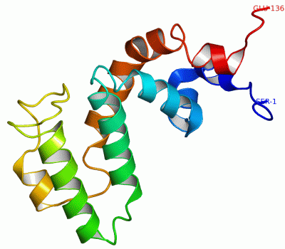
 Description
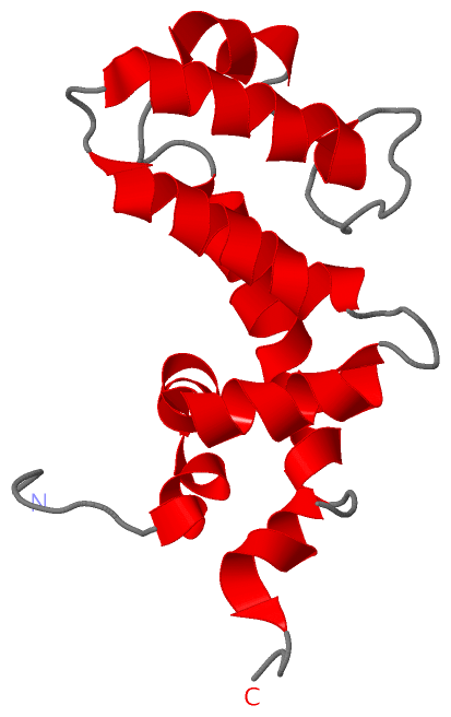
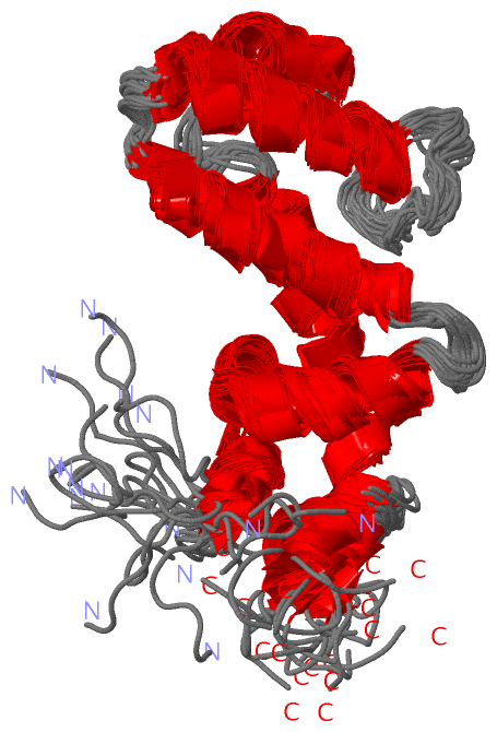