| molecular function |
|---|
| | GO:0003779 | | actin binding | | Interacting selectively and non-covalently with monomeric or multimeric forms of actin, including actin filaments. |
| | GO:0005509 | | calcium ion binding | | Interacting selectively and non-covalently with calcium ions (Ca2+). |
| | GO:0008092 | | cytoskeletal protein binding | | Interacting selectively and non-covalently with any protein component of any cytoskeleton (actin, microtubule, or intermediate filament cytoskeleton). |
| | GO:0046872 | | metal ion binding | | Interacting selectively and non-covalently with any metal ion. |
| | GO:0051010 | | microtubule plus-end binding | | Interacting selectively and non-covalently with the plus end of a microtubule. |
| | GO:0005515 | | protein binding | | Interacting selectively and non-covalently with any protein or protein complex (a complex of two or more proteins that may include other nonprotein molecules). |
| | GO:0042803 | | protein homodimerization activity | | Interacting selectively and non-covalently with an identical protein to form a homodimer. |
| biological process |
|---|
| | GO:0007409 | | axonogenesis | | De novo generation of a long process of a neuron, that carries efferent (outgoing) action potentials from the cell body towards target cells. Refers to the morphogenesis or creation of shape or form of the developing axon. |
| | GO:0007155 | | cell adhesion | | The attachment of a cell, either to another cell or to an underlying substrate such as the extracellular matrix, via cell adhesion molecules. |
| | GO:0007050 | | cell cycle arrest | | A regulatory process that halts progression through the cell cycle during one of the normal phases (G1, S, G2, M). |
| | GO:0007010 | | cytoskeleton organization | | A process that is carried out at the cellular level which results in the assembly, arrangement of constituent parts, or disassembly of cytoskeletal structures. |
| | GO:0045104 | | intermediate filament cytoskeleton organization | | A process that is carried out at the cellular level which results in the assembly, arrangement of constituent parts, or disassembly of cytoskeletal structures comprising intermediate filaments and their associated proteins. |
| | GO:0046907 | | intracellular transport | | The directed movement of substances within a cell. |
| | GO:0008090 | | retrograde axonal transport | | The directed movement of organelles or molecules along microtubules from the cell periphery toward the cell body in nerve cell axons. |
| cellular component |
|---|
| | GO:0031673 | | H zone | | A relatively pale zone traversing the center of the A band of a sarcomere, visible in relaxed muscle fibers; consists of the central portion of thick (myosin) filaments that are not overlapped by thin (actin) filaments. |
| | GO:0030018 | | Z disc | | Platelike region of a muscle sarcomere to which the plus ends of actin filaments are attached. |
| | GO:0015629 | | actin cytoskeleton | | The part of the cytoskeleton (the internal framework of a cell) composed of actin and associated proteins. Includes actin cytoskeleton-associated complexes. |
| | GO:0030424 | | axon | | The long process of a neuron that conducts nerve impulses, usually away from the cell body to the terminals and varicosities, which are sites of storage and release of neurotransmitter. |
| | GO:1904115 | | axon cytoplasm | | Any cytoplasm that is part of a axon. |
| | GO:0005938 | | cell cortex | | The region of a cell that lies just beneath the plasma membrane and often, but not always, contains a network of actin filaments and associated proteins. |
| | GO:0030054 | | cell junction | | A cellular component that forms a specialized region of connection between two or more cells or between a cell and the extracellular matrix. At a cell junction, anchoring proteins extend through the plasma membrane to link cytoskeletal proteins in one cell to cytoskeletal proteins in neighboring cells or to proteins in the extracellular matrix. |
| | GO:0042995 | | cell projection | | A prolongation or process extending from a cell, e.g. a flagellum or axon. |
| | GO:0005737 | | cytoplasm | | All of the contents of a cell excluding the plasma membrane and nucleus, but including other subcellular structures. |
| | GO:0009898 | | cytoplasmic side of plasma membrane | | The leaflet the plasma membrane that faces the cytoplasm and any proteins embedded or anchored in it or attached to its surface. |
| | GO:0005856 | | cytoskeleton | | Any of the various filamentous elements that form the internal framework of cells, and typically remain after treatment of the cells with mild detergent to remove membrane constituents and soluble components of the cytoplasm. The term embraces intermediate filaments, microfilaments, microtubules, the microtrabecular lattice, and other structures characterized by a polymeric filamentous nature and long-range order within the cell. The various elements of the cytoskeleton not only serve in the maintenance of cellular shape but also have roles in other cellular functions, including cellular movement, cell division, endocytosis, and movement of organelles. |
| | GO:0005783 | | endoplasmic reticulum | | The irregular network of unit membranes, visible only by electron microscopy, that occurs in the cytoplasm of many eukaryotic cells. The membranes form a complex meshwork of tubular channels, which are often expanded into slitlike cavities called cisternae. The ER takes two forms, rough (or granular), with ribosomes adhering to the outer surface, and smooth (with no ribosomes attached). |
| | GO:0005789 | | endoplasmic reticulum membrane | | The lipid bilayer surrounding the endoplasmic reticulum. |
| | GO:0005925 | | focal adhesion | | Small region on the surface of a cell that anchors the cell to the extracellular matrix and that forms a point of termination of actin filaments. |
| | GO:0030056 | | hemidesmosome | | A cell-substrate junction (attachment structure) found in epithelial cells that links intermediate filaments to extracellular matrices via transmembrane complexes. In vertebrates, hemidesmosomes mediate contact between the basal side of epithelial cells and the basal lamina. In C. elegans, hemidesmosomes connect epithelial cells to distinct extracellular matrices on both the apical and basal cell surfaces. |
| | GO:0016021 | | integral component of membrane | | The component of a membrane consisting of the gene products and protein complexes having at least some part of their peptide sequence embedded in the hydrophobic region of the membrane. |
| | GO:0014704 | | intercalated disc | | A complex cell-cell junction at which myofibrils terminate in cardiomyocytes; mediates mechanical and electrochemical integration between individual cardiomyocytes. The intercalated disc contains regions of tight mechanical attachment (fasciae adherentes and desmosomes) and electrical coupling (gap junctions) between adjacent cells. |
| | GO:0005882 | | intermediate filament | | A cytoskeletal structure that forms a distinct elongated structure, characteristically 10 nm in diameter, that occurs in the cytoplasm of eukaryotic cells. Intermediate filaments form a fibrous system, composed of chemically heterogeneous subunits and involved in mechanically integrating the various components of the cytoplasmic space. Intermediate filaments may be divided into five chemically distinct classes: Type I, acidic keratins; Type II, basic keratins; Type III, including desmin, vimentin and others; Type IV, neurofilaments and related filaments; and Type V, lamins. |
| | GO:0016020 | | membrane | | A lipid bilayer along with all the proteins and protein complexes embedded in it an attached to it. |
| | GO:0005874 | | microtubule | | Any of the long, generally straight, hollow tubes of internal diameter 12-15 nm and external diameter 24 nm found in a wide variety of eukaryotic cells; each consists (usually) of 13 protofilaments of polymeric tubulin, staggered in such a manner that the tubulin monomers are arranged in a helical pattern on the microtubular surface, and with the alpha/beta axes of the tubulin subunits parallel to the long axis of the tubule; exist in equilibrium with pool of tubulin monomers and can be rapidly assembled or disassembled in response to physiological stimuli; concerned with force generation, e.g. in the spindle. |
| | GO:0015630 | | microtubule cytoskeleton | | The part of the cytoskeleton (the internal framework of a cell) composed of microtubules and associated proteins. |
| | GO:0035371 | | microtubule plus-end | | The growing (plus) end of a microtubule. In vitro, microtubules polymerize more quickly at the plus end than at the minus end. In vivo, microtubule growth occurs only at the plus end, and the plus end switches between periods of growth and shortening, a behavior known as dynamic instability. |
| | GO:0005635 | | nuclear envelope | | The double lipid bilayer enclosing the nucleus and separating its contents from the rest of the cytoplasm; includes the intermembrane space, a gap of width 20-40 nm (also called the perinuclear space). |
| | GO:0005634 | | nucleus | | A membrane-bounded organelle of eukaryotic cells in which chromosomes are housed and replicated. In most cells, the nucleus contains all of the cell's chromosomes except the organellar chromosomes, and is the site of RNA synthesis and processing. In some species, or in specialized cell types, RNA metabolism or DNA replication may be absent. |
| | GO:0097038 | | perinuclear endoplasmic reticulum | | The portion of endoplasmic reticulum, the intracellular network of tubules and cisternae, that occurs near the nucleus. The lumen of the perinuclear endoplasmic reticulum is contiguous with the nuclear envelope lumen (also called perinuclear space), the region between the inner and outer nuclear membranes. |
| | GO:0048471 | | perinuclear region of cytoplasm | | Cytoplasm situated near, or occurring around, the nucleus. |
| | GO:0005886 | | plasma membrane | | The membrane surrounding a cell that separates the cell from its external environment. It consists of a phospholipid bilayer and associated proteins. |
| | GO:0042383 | | sarcolemma | | The outer membrane of a muscle cell, consisting of the plasma membrane, a covering basement membrane (about 100 nm thick and sometimes common to more than one fiber), and the associated loose network of collagen fibers. |
| | GO:0001725 | | stress fiber | | A contractile actin filament bundle that consists of short actin filaments with alternating polarity, cross-linked by alpha-actinin and possibly other actin bundling proteins, and with myosin present in a periodic distribution along the fiber. |
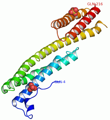
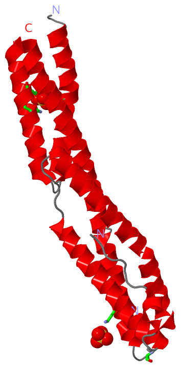
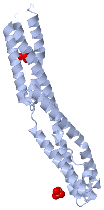
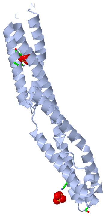
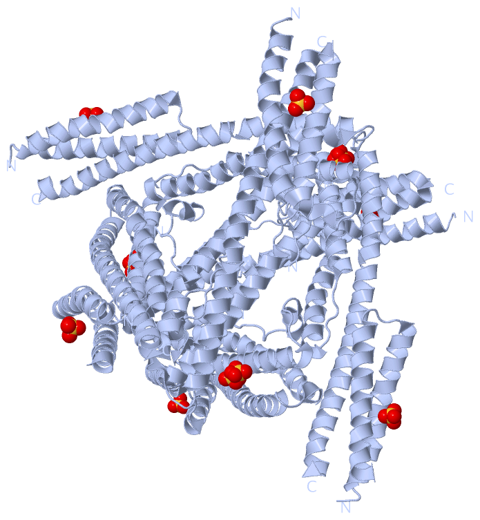
 Description
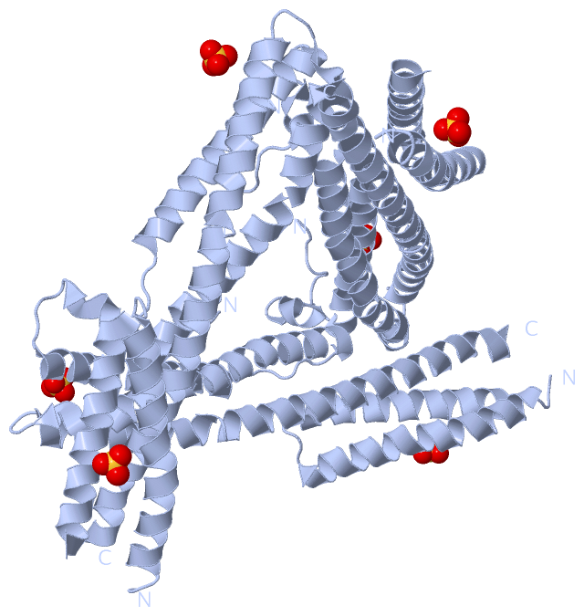