|
|
|
|
 Description
|
|
Compounds
|
||||||||||||||||||||||||||||||||||||||||||||||||||||
Chains, Units
Summary Information (see also Sequences/Alignments below) |
Ligands, Modified Residues, Ions (2, 5)| Asymmetric/Biological Unit (2, 5) |
Sites (1, 1)
Asymmetric Unit (1, 1)
|
SS Bonds (0, 0)| (no "SS Bond" information available for 2IAF) |
Cis Peptide Bonds (1, 1)
Asymmetric/Biological Unit
|
||||||||
SAPs(SNPs)/Variants (0, 0)| (no "SAP(SNP)/Variant" information available for 2IAF) |
PROSITE Motifs (0, 0)| (no "PROSITE Motif" information available for 2IAF) |
Exons (0, 0)| (no "Exon" information available for 2IAF) |
Sequences/Alignments
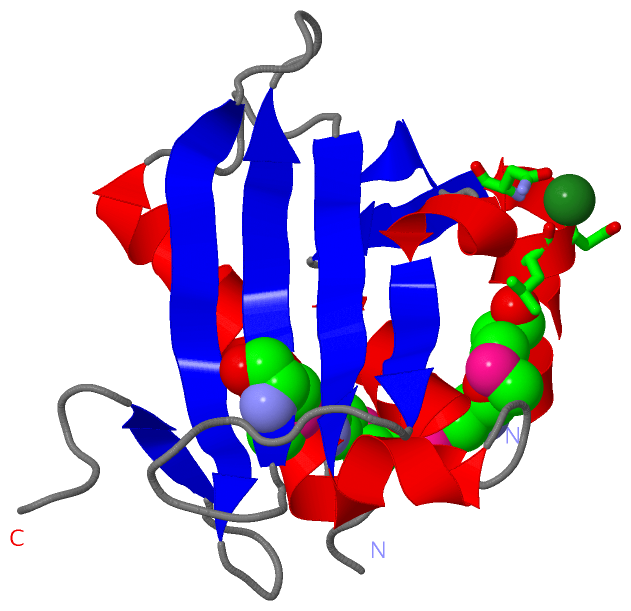
Asymmetric/Biological UnitChain A from PDB Type:PROTEIN Length:140 aligned with Q5WYB1_LEGPL | Q5WYB1 from UniProtKB/TrEMBL Length:458 Alignment length:145 26 36 46 56 66 76 86 96 106 116 126 136 146 156 Q5WYB1_LEGPL 17 SSHTVGPMLAANAFLQLLEQKNLFDKTQRVKVELYGSLALTGKGHGTDKAILNGLENKAPETVDPASMIPRMHEILDSNLLNLAGKKEIPFHEATDFLFLQKELLPKHSNGMRFSAFDGNANLLIEQVYYSIGGGFITTEEDFDK 161 SCOP domains d2iafa1 A:4-148 L-serine dehydratase SdhL, N-terminal domain SCOP domains CATH domains 2iafA00 A:4-148 D-3-phosphoglycerate dehydrogenase; domain 3 CATH domains Pfam domains ------------------------------------------------------------------------------------------------------------------------------------------------- Pfam domains SAPs(SNPs) ------------------------------------------------------------------------------------------------------------------------------------------------- SAPs(SNPs) PROSITE ------------------------------------------------------------------------------------------------------------------------------------------------- PROSITE Transcript ------------------------------------------------------------------------------------------------------------------------------------------------- Transcript 2iaf A 4 SSHTVGPmLAANAFLQLLEQKNLFDKTQRVKVELYGSLALTGKGHGTDKAILNGLENKAPE-----SmIPRmHEILDSNLLNLAGKKEIPFHEATDFLFLQKELLPKHSNGmRFSAFDGNANLLIEQVYYSIGGGFITTEEDFDK 148 |13 23 33 43 53 63| ||73 | 83 93 103 113 | 123 133 143 11-MSE 64 70| | 115-MSE 71-MSE 75-MSE
|
||||||||||||||||||||
SCOP Domains (1, 1)
Asymmetric/Biological Unit
 |
CATH Domains (1, 1)
Asymmetric/Biological Unit
|
Pfam Domains (0, 0)| (no "Pfam Domain" information available for 2IAF) |
Gene Ontology (4, 4)|
Asymmetric/Biological Unit(hide GO term definitions) Chain A (Q5WYB1_LEGPL | Q5WYB1)
|
||||||||||||||||||||||||||||||||||||
Interactive Views
|
||||||||||||||||||||||||||||||||||||||||||||||||||||||||||||||||||||||||||||||||||||||||||||||||||||||||||||||||||||||||||||||
Still Images
|
||||||||||||||||
Databases
|
||||||||||||||||||||||||||||||||||||||||||||||||||||||||||||||||||||||||||||||||||||||||||||||||||||||||||||||||||||||||||||||||||||||||||||||||||||||||||||||||
Analysis Tools
|
|||||||||||||||||||||||||||||||||||||||||||||||||||||||||||||
Entries Sharing at Least One Protein Chain (UniProt ID)
Related Entries Specified in the PDB File
|
|