|
|
|
|
 Description
|
|
Compounds
|
||||||||||||||||||||||||||||||||||||||||||||||||
Chains, Units
Summary Information (see also Sequences/Alignments below) |
Ligands, Modified Residues, Ions (3, 5)
Asymmetric Unit (3, 5)
|
Sites (5, 5)
Asymmetric Unit (5, 5)
|
SS Bonds (0, 0)| (no "SS Bond" information available for 2I7F) |
Cis Peptide Bonds (2, 2)
Asymmetric Unit
|
||||||||||||
SAPs(SNPs)/Variants (0, 0)| (no "SAP(SNP)/Variant" information available for 2I7F) |
PROSITE Motifs (0, 0)| (no "PROSITE Motif" information available for 2I7F) |
Exons (0, 0)| (no "Exon" information available for 2I7F) |
Sequences/Alignments
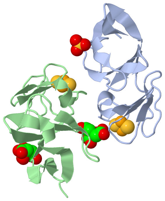
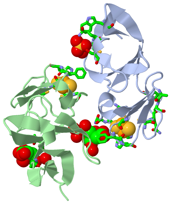
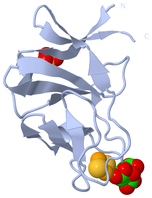
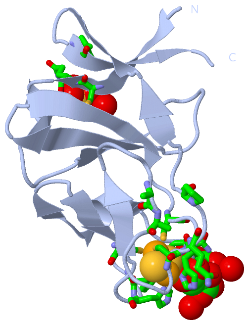
Asymmetric UnitChain A from PDB Type:PROTEIN Length:102 aligned with A2TC31_SPHYA | A2TC31 from UniProtKB/TrEMBL Length:108 Alignment length:102 12 22 32 42 52 62 72 82 92 102 A2TC31_SPHYA 3 NKLRLCQVASVKDGEPVAVYQEKMPALAVYNVDGEVFVTDNLCTHGNAMLTDGYQDGTIIECPFHGGSFDIATGAAKAFPCQIPIKTYPVTIEDGWVCIDQP 104 SCOP domains d2i7fa_ A: automated matches SCOP domains CATH domains 2i7fA00 A:3-104 'Rieske'-like iron-sulphur domains CATH domains Pfam domains ------------------------------------------------------------------------------------------------------ Pfam domains SAPs(SNPs) ------------------------------------------------------------------------------------------------------ SAPs(SNPs) PROSITE ------------------------------------------------------------------------------------------------------ PROSITE Transcript ------------------------------------------------------------------------------------------------------ Transcript 2i7f A 3 NKLRLCQVASVKDGEPVAVYQEKMPALAVYNVDGEVFVTDNLCTHGNAMLTDGYQDGTIIECPFHGGSFDIATGAAKAFPCQIPIKTYPVTIEDGWVCIDQP 104 12 22 32 42 52 62 72 82 92 102 Chain B from PDB Type:PROTEIN Length:102 aligned with A2TC31_SPHYA | A2TC31 from UniProtKB/TrEMBL Length:108 Alignment length:102 12 22 32 42 52 62 72 82 92 102 A2TC31_SPHYA 3 NKLRLCQVASVKDGEPVAVYQEKMPALAVYNVDGEVFVTDNLCTHGNAMLTDGYQDGTIIECPFHGGSFDIATGAAKAFPCQIPIKTYPVTIEDGWVCIDQP 104 SCOP domains d2i7fb_ B: automated matches SCOP domains CATH domains 2i7fB00 B:3-104 'Rieske'-like iron-sulphur domains CATH domains Pfam domains ------------------------------------------------------------------------------------------------------ Pfam domains SAPs(SNPs) ------------------------------------------------------------------------------------------------------ SAPs(SNPs) PROSITE ------------------------------------------------------------------------------------------------------ PROSITE Transcript ------------------------------------------------------------------------------------------------------ Transcript 2i7f B 3 NKLRLCQVASVKDGEPVAVYQEKMPALAVYNVDGEVFVTDNLCTHGNAMLTDGYQDGTIIECPFHGGSFDIATGAAKAFPCQIPIKTYPVTIEDGWVCIDQP 104 12 22 32 42 52 62 72 82 92 102
|
||||||||||||||||||||
SCOP Domains (1, 2)
Asymmetric Unit
 |
CATH Domains (1, 2)
Asymmetric Unit
|
Pfam Domains (0, 0)| (no "Pfam Domain" information available for 2I7F) |
Gene Ontology (6, 6)|
Asymmetric Unit(hide GO term definitions) Chain A,B (A2TC31_SPHYA | A2TC31)
|
||||||||||||||||||||||||||||||||||||||||||||||||
Interactive Views
|
|||||||||||||||||||||||||||||||||||||||||||||||||||||||||||||||||||||||||||||||||||||||||||||||||||||||||||||||||||||||||||||||||||||||||||||||||||||||||||||||||||||||||||||||||||||||||||||||
Still Images
|
||||||||||||||||
Databases
|
||||||||||||||||||||||||||||||||||||||||||||||||||||||||||||||||||||||||||||||||||||||||||||||||||||||||||||||||||||||||||||||||||||||||||||||||||||||||||||||||
Analysis Tools
|
|||||||||||||||||||||||||||||||||||||||||||||||||||||||||||||
Entries Sharing at Least One Protein Chain (UniProt ID)
Related Entries Specified in the PDB File
|
|