|
|
|
|
 Description
|
|
Compounds
|
||||||||||||||||||||||||||||||||||||||||||||||||
Chains, Units
Summary Information (see also Sequences/Alignments below) |
Ligands, Modified Residues, Ions (0, 0)| (no "Ligand,Modified Residues,Ions" information available for 2I3E) |
Sites (0, 0)| (no "Site" information available for 2I3E) |
SS Bonds (0, 0)| (no "SS Bond" information available for 2I3E) |
Cis Peptide Bonds (0, 0)| (no "Cis Peptide Bond" information available for 2I3E) |
SAPs(SNPs)/Variants (0, 0)| (no "SAP(SNP)/Variant" information available for 2I3E) |
PROSITE Motifs (0, 0)| (no "PROSITE Motif" information available for 2I3E) |
Exons (0, 0)| (no "Exon" information available for 2I3E) |
Sequences/Alignments
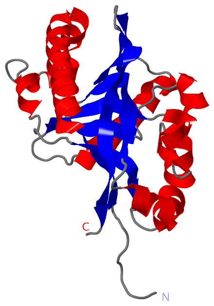
NMR StructureChain A from PDB Type:PROTEIN Length:222 aligned with Q90306_CARAU | Q90306 from UniProtKB/TrEMBL Length:411 Alignment length:222 177 187 197 207 217 227 237 247 257 267 277 287 297 307 317 327 337 347 357 367 377 387 Q90306_CARAU 168 QKEPELPLFFGWFLLPEEEERIKCATMDFLKTLDTLEAFKEHISEFTGEAEKEVDLEQYFQNPLQLHCTTKFCDYGKAEGAKEYAELQVVKESLTKSYELSVTALIVTPRTFGARVALTEAQVKLWPEGADKEGVAPALLPSVEALPAGSRAHVTLGCSAGVETVQTGLDLLEILALQKEGKEGTQVEMDLGTLTYLSEGRWFLALREPINADTTFTSFSED 389 SCOP domains d2i3ea_ A: automated matches SCOP domains CATH domains ------------------------------------------------------------------------------------------------------------------------------------------------------------------------------------------------------------------------------ CATH domains Pfam domains ------------------------------------------------------------------------------------------------------------------------------------------------------------------------------------------------------------------------------ Pfam domains SAPs(SNPs) ------------------------------------------------------------------------------------------------------------------------------------------------------------------------------------------------------------------------------ SAPs(SNPs) PROSITE ------------------------------------------------------------------------------------------------------------------------------------------------------------------------------------------------------------------------------ PROSITE Transcript ------------------------------------------------------------------------------------------------------------------------------------------------------------------------------------------------------------------------------ Transcript 2i3e A 1 GSHMELPLFFGWFLLPEEEERIKCATMDFLKTLDTLEAFKEHISEFTGEAEKEVDLEQYFQNPLQLHCTTKFCDYGKAEGAKEYAELQVVKESLTKSYELSVTALIVTPRTFGARVALTEAQVKLWPEGADKEGVAPALLPSVEALPAGSRAHVTLGCSAGVETVQTGLDLLEILALQKEGKEGTQVEMDLGTLTYLSEGRWFLALREPINADTTFTSFSED 222 10 20 30 40 50 60 70 80 90 100 110 120 130 140 150 160 170 180 190 200 210 220
|
||||||||||||||||||||
SCOP Domains (1, 1)
NMR Structure
 |
CATH Domains (0, 0)| (no "CATH Domain" information available for 2I3E) |
Pfam Domains (0, 0)| (no "Pfam Domain" information available for 2I3E) |
Gene Ontology (3, 3)|
NMR Structure(hide GO term definitions) Chain A (Q90306_CARAU | Q90306)
|
||||||||||||||||||||||||||||||||||||
Interactive Views
|
||||||||||||||||||||||||||||||||||||||||||||||||||||||||||||||||||||||||||||||||||||||||||||||||||||||||||||||||||||
Still Images
|
||||||||||||||||
Databases
|
||||||||||||||||||||||||||||||||||||||||||||||||||||||||||||||||||||||||||||||||||||||||||||||||||||||||||||||||||||||||||||||||||||||||||||||||||||||||||||||||
Analysis Tools
|
|||||||||||||||||||||||||||||||||||||||||||||||||||||||||||||
Entries Sharing at Least One Protein Chain (UniProt ID)
Related Entries Specified in the PDB File
|
|