| No. | Name | Evidence | Residues | Description |
|---|
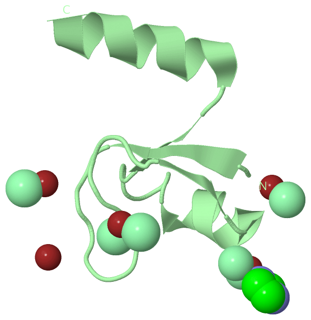
| 01 | AC1 | SOFTWARE | MET A:1 , ASP A:23 , CL A:203 , HOH A:210 , HOH B:306 | BINDING SITE FOR RESIDUE ZN A 101 |
| 02 | AC2 | SOFTWARE | ASP A:8 , LYS A:10 , CL A:204 , CL A:205 , HOH A:234 , HOH B:304 | BINDING SITE FOR RESIDUE ZN A 102 |
| 03 | AC3 | SOFTWARE | HOH A:220 , MET B:1 , ASP B:23 , CL B:210 | BINDING SITE FOR RESIDUE ZN B 103 |
| 04 | AC4 | SOFTWARE | ASP B:8 , CL B:206 , HOH B:309 , HOH B:322 | BINDING SITE FOR RESIDUE ZN B 104 |
| 05 | AC5 | SOFTWARE | GLU B:17 , CL B:201 , CL B:202 , HOH B:303 | BINDING SITE FOR RESIDUE ZN B 105 |
| 06 | AC6 | SOFTWARE | CL A:208 , HOH A:217 , HOH A:233 , HOH B:305 | BINDING SITE FOR RESIDUE ZN A 106 |
| 07 | AC7 | SOFTWARE | CL B:207 , CL B:209 , IMD B:301 , HOH B:308 | BINDING SITE FOR RESIDUE ZN B 107 |
| 08 | AC8 | SOFTWARE | HOH B:317 , HOH B:334 | BINDING SITE FOR RESIDUE ZN B 108 |
| 09 | AC9 | SOFTWARE | GLU B:17 , LYS B:19 , ZN B:105 , CL B:202 | BINDING SITE FOR RESIDUE CL B 201 |
| 10 | BC1 | SOFTWARE | GLY B:13 , LYS B:14 , GLU B:17 , ZN B:105 , CL B:201 | BINDING SITE FOR RESIDUE CL B 202 |
| 11 | BC2 | SOFTWARE | MET A:1 , ASP A:23 , ZN A:101 , HOH A:241 , LYS B:10 , HOH B:306 | BINDING SITE FOR RESIDUE CL A 203 |
| 12 | BC3 | SOFTWARE | ASP A:8 , LYS A:10 , ZN A:102 , CL A:205 , LYS B:14 , ASN B:20 | BINDING SITE FOR RESIDUE CL A 204 |
| 13 | BC4 | SOFTWARE | ASP A:8 , ZN A:102 , CL A:204 , HOH A:209 , HOH A:234 | BINDING SITE FOR RESIDUE CL A 205 |
| 14 | BC5 | SOFTWARE | ASN A:20 , ASP B:8 , ZN B:104 | BINDING SITE FOR RESIDUE CL B 206 |
| 15 | BC6 | SOFTWARE | ALA B:22 , TYR B:25 , ZN B:107 , CL B:209 , IMD B:301 | BINDING SITE FOR RESIDUE CL B 207 |
| 16 | BC7 | SOFTWARE | ALA A:22 , TYR A:25 , ZN A:106 | BINDING SITE FOR RESIDUE CL A 208 |
| 17 | BC8 | SOFTWARE | ZN B:107 , CL B:207 , IMD B:301 | BINDING SITE FOR RESIDUE CL B 209 |
| 18 | BC9 | SOFTWARE | ASP A:8 , MET B:1 , ASP B:23 , ZN B:103 | BINDING SITE FOR RESIDUE CL B 210 |
| 19 | CC1 | SOFTWARE | ASP A:8 , ASP B:23 , ZN B:107 , CL B:207 , CL B:209 | BINDING SITE FOR RESIDUE IMD B 301 |
 Description