|
|
|
|
 Description
|
|
Compounds
|
||||||||||||||||||||||||||||||||||||||||||||||||||||||||||||||||||||||||||
Chains, Units
Summary Information (see also Sequences/Alignments below) |
Ligands, Modified Residues, Ions (3, 6)
Asymmetric Unit (3, 6)
|
Sites (6, 6)
Asymmetric Unit (6, 6)
|
SS Bonds (0, 0)| (no "SS Bond" information available for 2H1C) |
Cis Peptide Bonds (0, 0)| (no "Cis Peptide Bond" information available for 2H1C) |
SAPs(SNPs)/Variants (0, 0)| (no "SAP(SNP)/Variant" information available for 2H1C) |
PROSITE Motifs (0, 0)| (no "PROSITE Motif" information available for 2H1C) |
Exons (0, 0)| (no "Exon" information available for 2H1C) |
Sequences/Alignments
Asymmetric UnitChain A from PDB Type:PROTEIN Length:139 aligned with FITB_NEIG1 | Q5F882 from UniProtKB/Swiss-Prot Length:139 Alignment length:139 10 20 30 40 50 60 70 80 90 100 110 120 130 FITB_NEIG1 1 MILLDTNVISEPLRPQPNERVVAWLDSLILEDVYLSAITVAELRLGVALLLNGKKKNVLHERLEQSILPLFAGRILPFDEPVAAIYAQIRSYAKTHGKEIAAADGYIAATAKQHSLTVATRDTGSFFAADVAVFNPWHD 139 SCOP domains d2h1ca1 A:1-138 Trafficking protein B - SCOP domains CATH domains ------------------------------------------------------------------------------------------------------------------------------------------- CATH domains Pfam domains ------------------------------------------------------------------------------------------------------------------------------------------- Pfam domains SAPs(SNPs) ------------------------------------------------------------------------------------------------------------------------------------------- SAPs(SNPs) PROSITE ------------------------------------------------------------------------------------------------------------------------------------------- PROSITE Transcript ------------------------------------------------------------------------------------------------------------------------------------------- Transcript 2h1c A 1 MILLDTNVISEPLRPQPNERVVAWLDSLILEDVYLSAITVAELRLGVALLLNGKKKNVLHERLEQSILPLFAGRILPFDEPVAAIYAQIRSYAKTHGKEIAAADGYIAATAKQHSLTVATRDTGSFFAADVAVFNPWHL 139 10 20 30 40 50 60 70 80 90 100 110 120 130 Chain B from PDB Type:PROTEIN Length:19 aligned with FITA_NEIG1 | Q5F881 from UniProtKB/Swiss-Prot Length:78 Alignment length:19 55 FITA_NEIG1 46 VRLGSMLASIGQEIGGVEL 64 SCOP domains ------------------- SCOP domains CATH domains ------------------- CATH domains Pfam domains ------------------- Pfam domains SAPs(SNPs) ------------------- SAPs(SNPs) PROSITE ------------------- PROSITE Transcript ------------------- Transcript 2h1c B 46 VRLGSMLASIGQEIGGVEL 64 55
|
||||||||||||||||||||
SCOP Domains (1, 1)
Asymmetric Unit

|
CATH Domains (0, 0)| (no "CATH Domain" information available for 2H1C) |
Pfam Domains (0, 0)| (no "Pfam Domain" information available for 2H1C) |
Gene Ontology (13, 16)|
Asymmetric Unit(hide GO term definitions) Chain A (FITB_NEIG1 | Q5F882)
Chain B (FITA_NEIG1 | Q5F881)
|
||||||||||||||||||||||||||||||||||||||||||||||||||||||||||||||||||||||||||||||||||||||||||||||||||||||||||||||||||||||||
Interactive Views
|
||||||||||||||||||||||||||||||||||||||||||||||||||||||||||||||||||||||||||||||||||||||||||||||||||||||||||||||||||||||||||||||||||||||||||||||||||||||||||||||||||||||||||||||||||||||||||||||
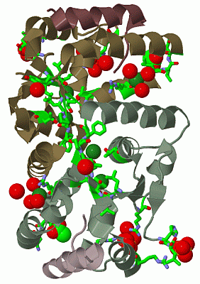
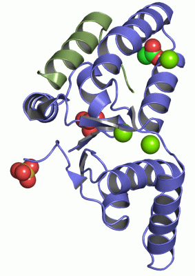
Still Images
|
||||||||||||||||
Databases
|
||||||||||||||||||||||||||||||||||||||||||||||||||||||||||||||||||||||||||||||||||||||||||||||||||||||||||||||||||||||||||||||||||||||||||||||||||||||||||||||||||||||||||||||||||||||||||
Analysis Tools
|
||||||||||||||||||||||||||||||||||||||||||||||||||||||||||||||||||||||||
Entries Sharing at Least One Protein Chain (UniProt ID)
Related Entries Specified in the PDB File
|
|