Show PDB file:   
         Plain Text   HTML   (compressed file size)
QuickSearch:   
by PDB,NDB,UniProt,PROSITE Code or Search Term(s)  
(-)Asym./Biol. Unit - manually
(-)Asym./Biol. Unit
(-)Asym./Biol. Unit - sites
collapse expand < >
Image Asym./Biol. Unit - manually
Asym./Biol. Unit - manually  (Jmol Viewer)
Image Asym./Biol. Unit
Asym./Biol. Unit  (Jmol Viewer)
Image Asym./Biol. Unit - sites
Asym./Biol. Unit - sites  (Jmol Viewer)

(-) Description

Title :  THE STRUCTURE OF THE ORTHORHOMBIC FORM OF SOFT-SHELLED TURTLE LYSOZYME AT 1.9 ANGSTROMS RESOLUTION
 
Authors :  J. Siritapetawee, S. Thammasirirak, J. Yuvaniyama, R. C. Robinson
Date :  02 May 06  (Deposition) - 08 May 07  (Release) - 24 Feb 09  (Revision)
Method :  X-RAY DIFFRACTION
Resolution :  1.90
Chains :  Asym./Biol. Unit :  A
Keywords :  Lysozyme, Hydrolase (Keyword Search: [Gene Ontology, PubMed, Web (Google))
 
Reference :  J. Siritapetawee, S. Thammasirirak, J. Yuvaniyama, R. C. Robinson
Crystallization And The 1. 9 Angstroms Of The Egg-White Lysozyme From A Taiwanese Soft-Shelled Turtle (Trionyx Sinensis Wiegmann)
To Be Published
PubMed: search
(for further references see the PDB file header)

(-) Compounds

Molecule 1 - LYSOZYME C
    ChainsA
    EC Number3.2.1.17
    Organism CommonCHINESE SOFTSHELL TURTLE
    Organism ScientificPELODISCUS SINENSIS
    Organism Taxid13735
    Other DetailsTHE CORRECT NAME OF THE SOURCE IS TRIONYX SINENSIS WIEGMANN
    Synonym1,4-BETA-N-ACETYLMURAMIDASE C

 Structural Features

(-) Chains, Units

  1
Asymmetric/Biological Unit A

Summary Information (see also Sequences/Alignments below)

(-) Ligands, Modified Residues, Ions  (1, 1)

Asymmetric/Biological Unit (1, 1)
No.NameCountTypeFull Name
1SO41Ligand/IonSULFATE ION

(-) Sites  (1, 1)

Asymmetric Unit (1, 1)
No.NameEvidenceResiduesDescription
1AC1SOFTWAREGLY A:1 , LYS A:87 , ALA A:88 , GLN A:124 , HOH A:159 , HOH A:185 , HOH A:195BINDING SITE FOR RESIDUE SO4 A 157

(-) SS Bonds  (4, 4)

Asymmetric/Biological Unit
No.Residues
1A:7 -A:129
2A:31 -A:117
3A:66 -A:82
4A:78 -A:96

(-) Cis Peptide Bonds  (0, 0)

(no "Cis Peptide Bond" information available for 2GV0)

 Sequence-Structure Mapping

(-) SAPs(SNPs)/Variants  (0, 0)

(no "SAP(SNP)/Variant" information available for 2GV0)

(-) PROSITE Motifs  (2, 2)

Asymmetric/Biological Unit (2, 2)
 PROSITEUniProtKBPDB
No.IDACDescriptionIDLocationCountLocation
1LACTALBUMIN_LYSOZYME_2PS51348 Alpha-lactalbumin / lysozyme C family profile.LYSC_PELSI2-131  1A:2-131
2LACTALBUMIN_LYSOZYME_1PS00128 Alpha-lactalbumin / lysozyme C signature.LYSC_PELSI78-96  1A:78-96

(-) Exons   (0, 0)

(no "Exon" information available for 2GV0)

(-) Sequences/Alignments

Asymmetric/Biological Unit
   Reformat: Number of residues per line =  ('0' or empty: single-line sequence representation)
  Number of residues per labelling interval =   
  UniProt sequence: complete  aligned part    
   Show mapping: SCOP domains CATH domains Pfam domains Secondary structure (by author)
SAPs(SNPs) PROSITE motifs Exons
(details for a mapped element are shown in a popup box when the mouse pointer rests over it)
Chain A from PDB  Type:PROTEIN  Length:131
 aligned with LYSC_PELSI | Q7LZQ1 from UniProtKB/Swiss-Prot  Length:131

    Alignment length:131
                                    10        20        30        40        50        60        70        80        90       100       110       120       130 
           LYSC_PELSI     1 GKIYEQCELAREFKRHGMDGYHGYSLGDWVCTAKHESNFNTAATNYNRGDQSTDYGILQINSRWWCNDGKTPKAKNACGIECSELLKADITAAVNCAKRIVRDPNGMGAWVAWTKYCKGKDVSQWIKGCKL 131
               SCOP domains d2gv0a_ A: automated matches                                                                                                        SCOP domains
               CATH domains 2gv0A00 A:1-131  [code=1.10.530.10, no name defined]                                                                                CATH domains
               Pfam domains ----------------------------------------------------------------------------------------------------------------------------------- Pfam domains
         Sec.struct. author ....hhhhhhhhhhhh...hhhhhhhhhhhhhhhhhhh.....eeee....eeee....ee...................hhhhhhh..hhhhhhhhhhhhhh.hhhhhhhhhhhhh....hhhhh..... Sec.struct. author
                 SAPs(SNPs) ----------------------------------------------------------------------------------------------------------------------------------- SAPs(SNPs)
                PROSITE (1) -LACTALBUMIN_LYSOZYME_2  PDB: A:2-131 UniProt: 2-131                                                                                PROSITE (1)
                PROSITE (2) -----------------------------------------------------------------------------LACTALBUMIN_LYSOZYM----------------------------------- PROSITE (2)
                 Transcript ----------------------------------------------------------------------------------------------------------------------------------- Transcript
                 2gv0 A   1 GKIYEQCELAREFKRHGMDGYHGYSLGDWVCTAKHESNFNTAATNYNRGDQSTDYGILQINSRWWCNDGKTPKAKNACGIECSELLKADITAAVNCAKRIVRDPNGMGAWVAWTKYCKGKDVSQWIKGCKL 131
                                    10        20        30        40        50        60        70        80        90       100       110       120       130 

   Legend:   → Mismatch (orange background)
  - → Gap (green background, '-', border residues have a numbering label)
    → Modified Residue (blue background, lower-case, 'x' indicates undefined single-letter code, labelled with number + name)
  x → Chemical Group (purple background, 'x', labelled with number + name, e.g. ACE or NH2)
  extra numbering lines below/above indicate numbering irregularities and modified residue names etc., number ends below/above '|'

 Classification and Annotation

(-) SCOP Domains  (1, 1)

Asymmetric/Biological Unit

(-) CATH Domains  (1, 1)

Asymmetric/Biological Unit

(-) Pfam Domains  (0, 0)

(no "Pfam Domain" information available for 2GV0)

(-) Gene Ontology  (8, 8)

Asymmetric/Biological Unit(hide GO term definitions)
Chain A   (LYSC_PELSI | Q7LZQ1)
molecular function
    GO:0003824    catalytic activity    Catalysis of a biochemical reaction at physiological temperatures. In biologically catalyzed reactions, the reactants are known as substrates, and the catalysts are naturally occurring macromolecular substances known as enzymes. Enzymes possess specific binding sites for substrates, and are usually composed wholly or largely of protein, but RNA that has catalytic activity (ribozyme) is often also regarded as enzymatic.
    GO:0016787    hydrolase activity    Catalysis of the hydrolysis of various bonds, e.g. C-O, C-N, C-C, phosphoric anhydride bonds, etc. Hydrolase is the systematic name for any enzyme of EC class 3.
    GO:0016798    hydrolase activity, acting on glycosyl bonds    Catalysis of the hydrolysis of any glycosyl bond.
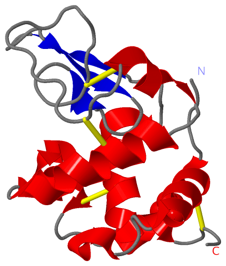
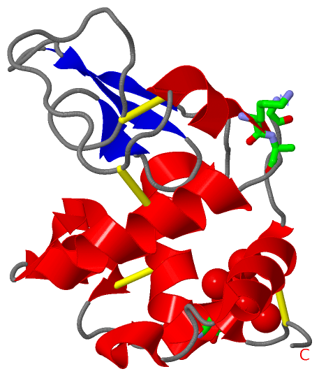
    GO:0003796    lysozyme activity    Catalysis of the hydrolysis of the beta-(1->4) linkages between N-acetylmuramic acid and N-acetyl-D-glucosamine residues in a peptidoglycan.
biological process
    GO:0019835    cytolysis    The rupture of cell membranes and the loss of cytoplasm.
    GO:0042742    defense response to bacterium    Reactions triggered in response to the presence of a bacterium that act to protect the cell or organism.
    GO:0008152    metabolic process    The chemical reactions and pathways, including anabolism and catabolism, by which living organisms transform chemical substances. Metabolic processes typically transform small molecules, but also include macromolecular processes such as DNA repair and replication, and protein synthesis and degradation.
cellular component
    GO:0005576    extracellular region    The space external to the outermost structure of a cell. For cells without external protective or external encapsulating structures this refers to space outside of the plasma membrane. This term covers the host cell environment outside an intracellular parasite.

 Visualization

(-) Interactive Views

Asymmetric/Biological Unit
  Complete Structure
    Jena3D(integrated viewing of ligand, site, SAP, PROSITE, SCOP information)
    WebMol | AstexViewer[tm]@PDBe
(Java Applets, require no local installation except for Java; loading may be slow)
    STRAP
(Java WebStart application, automatic local installation, requires Java; full application with system access!)
    RasMol
(require local installation)
    Molscript (VRML)
(requires installation of a VRML viewer; select preferred view via VRML and generate a mono or stereo PDF format file)
 
  Ligands, Modified Residues, Ions
    SO4  [ RasMol | Jena3D ]  +environment [ RasMol | Jena3D ]
 
  Sites
    AC1  [ RasMol ]  +environment [ RasMol ]
 
  Cis Peptide Bonds
(no "Cis Peptide Bonds" information available for 2gv0)
 

(-) Still Images

Jmol
  protein: cartoon or spacefill or dots and stick; nucleic acid: cartoon and stick; ligands: spacefill; active site: stick
Molscript
  protein, nucleic acid: cartoon; ligands: spacefill; active site: ball and stick

 Databases and Analysis Tools

(-) Databases

Access by PDB/NDB ID
  2gv0
    Family and Domain InformationProDom | SYSTERS
    General Structural InformationGlycoscienceDB | MMDB | NDB | OCA | PDB | PDBe | PDBj | PDBsum | PDBWiki | PQS | PROTEOPEDIA
    Orientation in MembranesOPM
    Protein SurfaceSURFACE
    Secondary StructureDSSP (structure derived) | HSSP (homology derived)
    Structural GenomicsGeneCensus
    Structural NeighboursCE | VAST
    Structure ClassificationCATH | Dali | SCOP
    Validation and Original DataBMRB Data View | BMRB Restraints Grid | EDS | PROCHECK | RECOORD | WHAT_CHECK
 
Access by UniProt ID/Accession number
  LYSC_PELSI | Q7LZQ1
    Comparative Protein Structure ModelsModBase
    Genomic InformationEnsembl
    Protein-protein InteractionDIP
    Sequence, Family and Domain InformationInterPro | Pfam | SMART | UniProtKB/SwissProt
 
Access by Enzyme Classificator   (EC Number)
  3.2.1.17
    General Enzyme InformationBRENDA | EC-PDB | Enzyme | IntEnz
    PathwayKEGG | MetaCyc
 
Access by Disease Identifier   (MIM ID)
  (no 'MIM ID' available)
    Disease InformationOMIM
 
Access by GenAge ID
  (no 'GenAge ID' available)
    Age Related InformationGenAge

(-) Analysis Tools

Access by PDB/NDB ID
    Domain InformationXDom
    Interatomic Contacts of Structural UnitsCSU
    Ligand-protein ContactsLPC
    Protein CavitiescastP
    Sequence and Secondary StructurePDBCartoon
    Structure AlignmentSTRAP(Java WebStart application, automatic local installation, requires Java; full application with system access!)
    Structure and Sequence BrowserSTING
 
Access by UniProt ID/Accession number
  LYSC_PELSI | Q7LZQ1
    Protein Disorder PredictionDisEMBL | FoldIndex | GLOBPLOT (for more information see DisProt)

 Related Entries

(-) Entries Sharing at Least One Protein Chain (UniProt ID)

(no "Entries Sharing at Least One Protein Chain" available for 2GV0)

(-) Related Entries Specified in the PDB File

(no "Related Entries Specified in the PDB File" available for 2GV0)