|
|
|
|
 Description
|
|
Compounds
|
||||||||||||||||||||||||||||||||||||||||
Chains, Units
Summary Information (see also Sequences/Alignments below) |
Ligands, Modified Residues, Ions (2, 4)| Asymmetric Unit (2, 4) Biological Unit 1 (2, 2) Biological Unit 2 (2, 2) |
Sites (4, 4)
Asymmetric Unit (4, 4)
|
SS Bonds (0, 0)| (no "SS Bond" information available for 2EXV) |
Cis Peptide Bonds (0, 0)| (no "Cis Peptide Bond" information available for 2EXV) |
SAPs(SNPs)/Variants (1, 2)
Asymmetric Unit (1, 2)
|
||||||||||||||||||||||||||||||||||||||||||||||||||||||||||||||||||||||||||||||||||||||||||||||||||||||||||||||||||||||||||||||||||||||||||||||||||||||||||||||||||||||||||||||
PROSITE Motifs (0, 0)| (no "PROSITE Motif" information available for 2EXV) |
Exons (0, 0)| (no "Exon" information available for 2EXV) |
Sequences/Alignments
Asymmetric UnitChain A from PDB Type:PROTEIN Length:82 aligned with CY551_PSEAE | P00099 from UniProtKB/Swiss-Prot Length:104 Alignment length:82 32 42 52 62 72 82 92 102 CY551_PSEAE 23 EDPEVLFKNKGCVACHAIDTKMVGPAYKDVAAKFAGQAGAEAELAQRIKNGSQGVWGPIPMPPNAVSDDEAQTLAKWVLSQK 104 SCOP domains d2exva_ A: automated matches SCOP domains CATH domains 2exvA00 A:1-82 Cytochrome c CATH domains Pfam domains ---------------------------------------------------------------------------------- Pfam domains SAPs(SNPs) -A-------------------------------------------------------------------------------- SAPs(SNPs) PROSITE ---------------------------------------------------------------------------------- PROSITE Transcript ---------------------------------------------------------------------------------- Transcript 2exv A 1 EDPEVLAKNKGCVACHAIDTKMVGPAYKDVAAKFAGQAGAEAELAQRIKNGSQGVWGPIPMPPNAVSDDEAQTLAKWVLSQK 82 10 20 30 40 50 60 70 80 Chain C from PDB Type:PROTEIN Length:81 aligned with CY551_PSEAE | P00099 from UniProtKB/Swiss-Prot Length:104 Alignment length:81 33 43 53 63 73 83 93 103 CY551_PSEAE 24 DPEVLFKNKGCVACHAIDTKMVGPAYKDVAAKFAGQAGAEAELAQRIKNGSQGVWGPIPMPPNAVSDDEAQTLAKWVLSQK 104 SCOP domains d2exvc_ C: automated matches SCOP domains CATH domains 2exvC00 C:2-82 Cytochrome c CATH domains Pfam domains --------------------------------------------------------------------------------- Pfam domains SAPs(SNPs) A-------------------------------------------------------------------------------- SAPs(SNPs) PROSITE --------------------------------------------------------------------------------- PROSITE Transcript --------------------------------------------------------------------------------- Transcript 2exv C 2 DPEVLAKNKGCVACHAIDTKMVGPAYKDVAAKFAGQAGAEAELAQRIKNGSQGVWGPIPMPPNAVSDDEAQTLAKWVLSQK 82 11 21 31 41 51 61 71 81
|
||||||||||||||||||||
SCOP Domains (1, 2)
Asymmetric Unit
 |
CATH Domains (1, 2)
Asymmetric Unit
|
Pfam Domains (0, 0)| (no "Pfam Domain" information available for 2EXV) |
Gene Ontology (6, 6)|
Asymmetric Unit(hide GO term definitions) Chain A,C (CY551_PSEAE | P00099)
|
||||||||||||||||||||||||||||||||||||||||||||||||||||||
Interactive Views
|
|||||||||||||||||||||||||||||||||||||||||||||||||||||||||||||||||||||||||||||||||||||||||||||||||||||||||||||||||||||||||||||||||||||||||||||||||||||||||||||||||||||||||
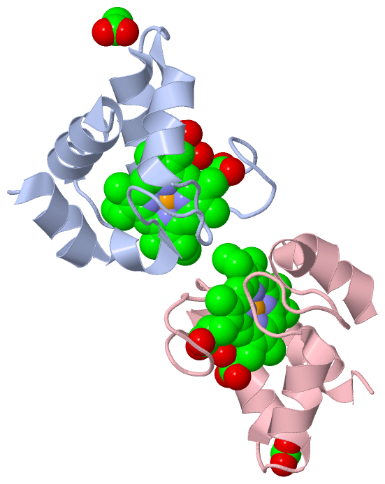
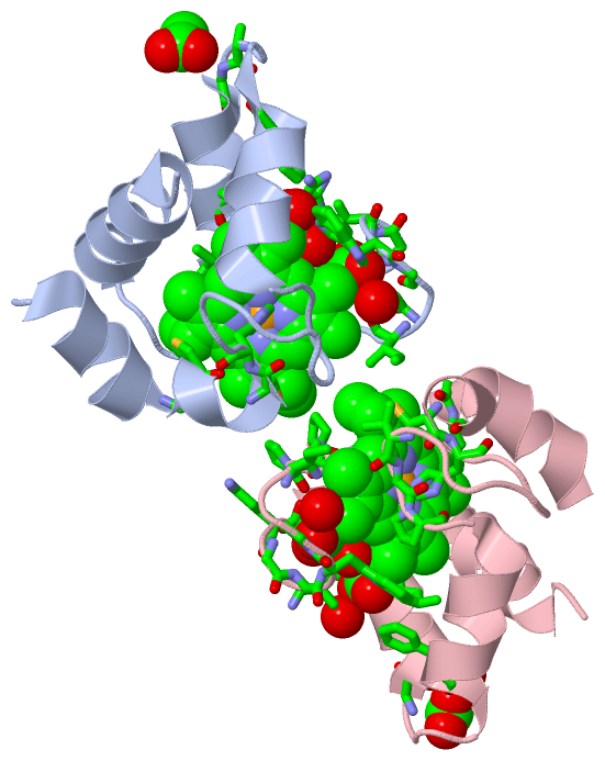
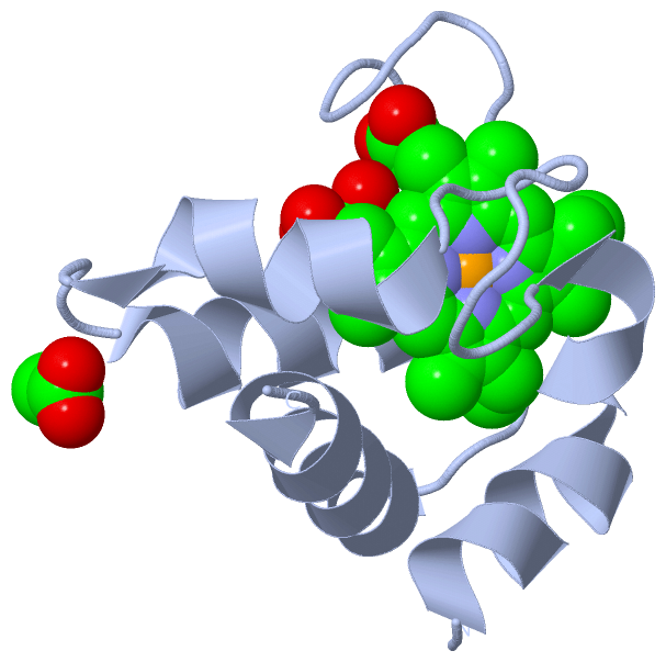
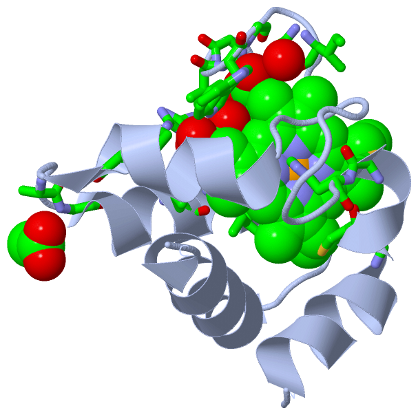
Still Images
|
||||||||||||||||
Databases
|
||||||||||||||||||||||||||||||||||||||||||||||||||||||||||||||||||||||||||||||||||||||||||||||||||||||||||||||||||||||||||||||||||||||||||||||||||||||||||||||||
Analysis Tools
|
|||||||||||||||||||||||||||||||||||||||||||||||||||||||||||||
Entries Sharing at Least One Protein Chain (UniProt ID)
Related Entries Specified in the PDB File
|
|