|
|
|
|
 Description
|
|
Compounds
|
||||||||||||||||||||||||||||||||||||||||
Chains, Units
Summary Information (see also Sequences/Alignments below) |
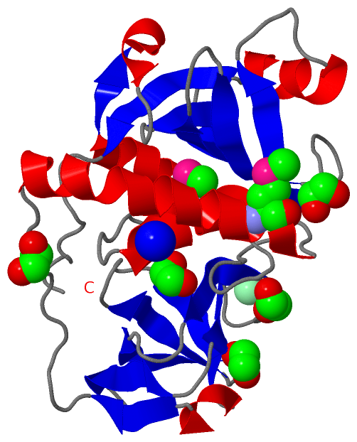
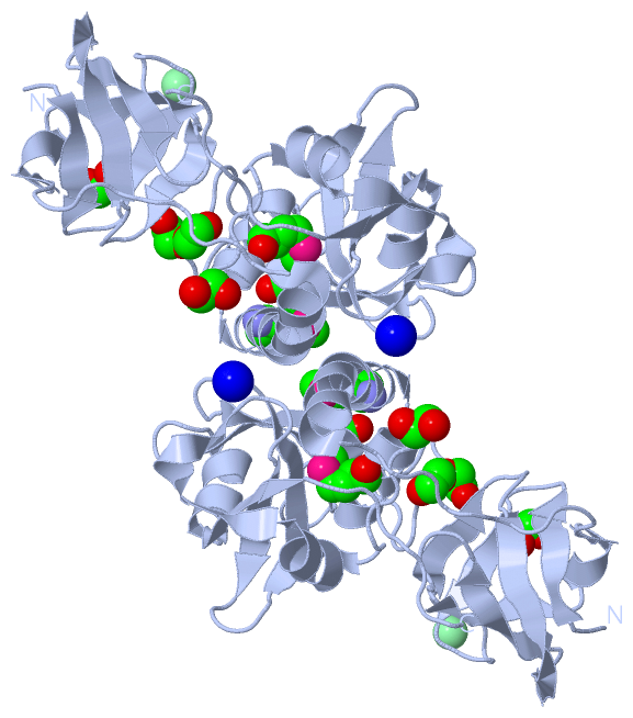
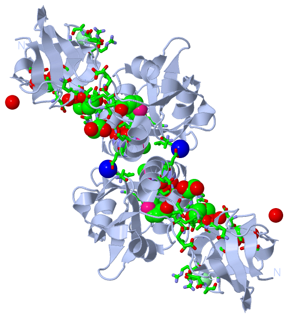
Ligands, Modified Residues, Ions (4, 9)| Asymmetric Unit (4, 9) Biological Unit 1 (2, 14) |
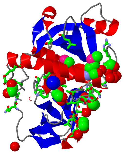
Sites (7, 7)
Asymmetric Unit (7, 7)
|
SS Bonds (0, 0)| (no "SS Bond" information available for 2EVR) |
Cis Peptide Bonds (2, 2)
Asymmetric Unit
|
||||||||||||
SAPs(SNPs)/Variants (0, 0)| (no "SAP(SNP)/Variant" information available for 2EVR) |
PROSITE Motifs (0, 0)| (no "PROSITE Motif" information available for 2EVR) |
Exons (0, 0)| (no "Exon" information available for 2EVR) |
Sequences/Alignments
Asymmetric UnitChain A from PDB Type:PROTEIN Length:222 aligned with B2J9B4_NOSP7 | B2J9B4 from UniProtKB/TrEMBL Length:242 Alignment length:222 30 40 50 60 70 80 90 100 110 120 130 140 150 160 170 180 190 200 210 220 230 240 B2J9B4_NOSP7 21 KLGEYQCLADLNLFDSPECTRLATQSASGRHLWVTSNHQNLAVEVYLCEDDYPGWLSLSDFDSLQPATVPYQAATFSESEIKKLLAEVIAFTQKAMQQSNYYLWGGTVGPNYDCSGLMQAAFASVGIWLPRDAYQQEGFTQPITIAELVAGDLVFFGTSQKATHVGLYLADGYYIHSSGKDQGRDGIGIDILSEQGDAVSLSYYQQLRGAGRVFKSYEPQRR 242 SCOP domains d2evra1 A:13-86 Cell wall-associated hydrolase Spr N-terminal domain d2evra2 A:87-234 Cell wall-associated hydrolase Spr C-terminal domain SCOP domains CATH domains 2evrA01 A:13-86 SH3 Domains 2evrA02 A:87-234 endopeptidase domain like (from Nostoc punctiforme) CATH domains Pfam domains ------------------------------------------------------------------------------------------------------------------------------------------------------------------------------------------------------------------------------ Pfam domains SAPs(SNPs) ------------------------------------------------------------------------------------------------------------------------------------------------------------------------------------------------------------------------------ SAPs(SNPs) PROSITE ------------------------------------------------------------------------------------------------------------------------------------------------------------------------------------------------------------------------------ PROSITE Transcript ------------------------------------------------------------------------------------------------------------------------------------------------------------------------------------------------------------------------------ Transcript 2evr A 13 KLGEYQCLADLNLFDSPECTRLATQSASGRHLWVTSNHQNLAVEVYLCEDDYPGWLSLSDFDSLQPATVPYQAATFSESEIKKLLAEVIAFTQKAmQQSNYYLWGGTVGPNYDCSGLmQAAFASVGIWLPRDAYQQEGFTQPITIAELVAGDLVFFGTSQKATHVGLYLADGYYIHSSGKDQGRDGIGIDILSEQGDAVSLSYYQQLRGAGRVFKSYEPQRR 234 22 32 42 52 62 72 82 92 102 | 112 122 132 142 152 162 172 182 192 202 212 222 232 108-MSE 130-MSE
|
||||||||||||||||||||
SCOP Domains (2, 2)
Asymmetric Unit
 |
CATH Domains (2, 2)
Asymmetric Unit
|
Pfam Domains (0, 0)| (no "Pfam Domain" information available for 2EVR) |
Gene Ontology (0, 0)|
Asymmetric Unit(hide GO term definitions)
(no "Gene Ontology" information available for 2EVR)
|
Interactive Views
|
|||||||||||||||||||||||||||||||||||||||||||||||||||||||||||||||||||||||||||||||||||||||||||||||||||||||||||||||||||||||||||||||||||||||||||||||||||||||||||||||||||||||||||||||||||||||||||||||||||||||||||||||
Still Images
|
||||||||||||||||
Databases
|
||||||||||||||||||||||||||||||||||||||||||||||||||||||||||||||||||||||||||||||||||||||||||||||||||||||||||||||||||||||||||||||||||||||||||||||||||||||||||||||||
Analysis Tools
|
|||||||||||||||||||||||||||||||||||||||||||||||||||||||||||||
Entries Sharing at Least One Protein Chain (UniProt ID)
Related Entries Specified in the PDB File
|
|