Show PDB file:   
         Plain Text   HTML   (compressed file size)
QuickSearch:   
by PDB,NDB,UniProt,PROSITE Code or Search Term(s)  
(-)NMR Structure - model 1
(-)NMR Structure - all models
collapse expand < >
Image NMR Structure - model 1
NMR Structure - model 1  (Jmol Viewer)
Image NMR Structure - all models
NMR Structure - all models  (Jmol Viewer)

(-) Description

Title :  SOLUTION STRUCTURE OF THE PH DOMAIN OF PLECKSTRIN HOMOLOGY DOMAIN-CONTAINING PROTEIN FAMILY B MEMBER 1 FROM MOUSE
 
Authors :  H. Li, T. Tomizawa, S. Koshiba, M. Inoue, T. Kigawa, S. Yokoyama, Riken Structural Genomics/Proteomics Initiative (Rsgi)
Date :  13 Dec 05  (Deposition) - 13 Jun 06  (Release) - 24 Feb 09  (Revision)
Method :  SOLUTION NMR
Resolution :  NOT APPLICABLE
Chains :  NMR Structure  :  A  (20x)
Keywords :  Ph Domain, Phret1, Structural Genomics, Nppsfa, National Project On Protein Structural And Functional Analyses, Riken Structural Genomics/Proteomics Initiative, Rsgi, Membrane Protein (Keyword Search: [Gene Ontology, PubMed, Web (Google))
 
Reference :  H. Li, T. Tomizawa, S. Koshiba, M. Inoue, T. Kigawa, S. Yokoyama
Solution Structure Of The Ph Domain Of Pleckstrin Homology Domain-Containing Protein Family B Member 1 From Mouse
To Be Published
PubMed: search
(for further references see the PDB file header)

(-) Compounds

Molecule 1 - PLECKSTRIN HOMOLOGY DOMAIN-CONTAINING PROTEIN FAMILY B MEMBER 1
    ChainsA
    EngineeredYES
    Expression System PlasmidP050302-02
    Expression System Vector TypePLASMID
    FragmentPH DOMAIN
    GenePHR1
    Organism CommonHOUSE MOUSE
    Organism ScientificMUS MUSCULUS
    Organism Taxid10090
    Other DetailsCELL-FREE PROTEIN SYNTHESIS
    SynonymPLECKSTRIN HOMOLOGY DOMAIN RETINAL PROTEIN 1, PH DOMAIN CONTAINING PROTEIN IN RETINA 1, PHRET1

 Structural Features

(-) Chains, Units

  
NMR Structure (20x)

Summary Information (see also Sequences/Alignments below)

(-) Ligands, Modified Residues, Ions  (0, 0)

(no "Ligand,Modified Residues,Ions" information available for 2D9V)

(-) Sites  (0, 0)

(no "Site" information available for 2D9V)

(-) SS Bonds  (0, 0)

(no "SS Bond" information available for 2D9V)

(-) Cis Peptide Bonds  (0, 0)

(no "Cis Peptide Bond" information available for 2D9V)

 Sequence-Structure Mapping

(-) SAPs(SNPs)/Variants  (0, 0)

(no "SAP(SNP)/Variant" information available for 2D9V)

(-) PROSITE Motifs  (1, 1)

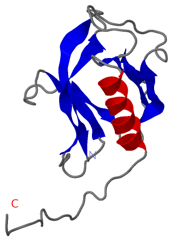
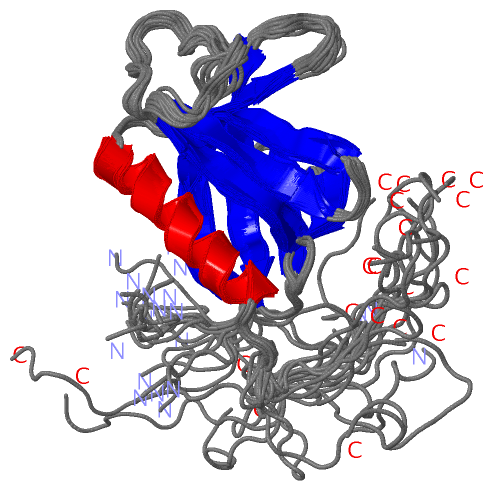
NMR Structure (1, 1)
 PROSITEUniProtKBPDB
No.IDACDescriptionIDLocationCountLocation
1PH_DOMAINPS50003 PH domain profile.PKHB1_MOUSE21-128  1A:8-114

(-) Exons   (0, 0)

(no "Exon" information available for 2D9V)

(-) Sequences/Alignments

NMR Structure
   Reformat: Number of residues per line =  ('0' or empty: single-line sequence representation)
  Number of residues per labelling interval =   
  UniProt sequence: complete  aligned part    
   Show mapping: SCOP domains CATH domains Pfam domains Secondary structure (by author)
SAPs(SNPs) PROSITE motifs Exons
(details for a mapped element are shown in a popup box when the mouse pointer rests over it)
Chain A from PDB  Type:PROTEIN  Length:130
 aligned with PKHB1_MOUSE | Q9QYE9 from UniProtKB/Swiss-Prot  Length:243

    Alignment length:207
                                    19        29        39        49        59        69        79        89        99       109       119       129       139       149       159       169       179       189       199       209       
          PKHB1_MOUSE    10 DSILESPFEEMALVRGGWLWRQSSILRRWKRNWFALWLDGTLGYYHDETAQDEEDRVVIHFNVRDIKVGQECQDVQPPEGRSRDGLLTVNLREGSRLHLCAETRDDAIAWKTALMEANSTPAPAGATVPPRSRRVCPKVRCTTLSWNPCKVERRIWVRVYSPYQDYYEVVPPNAHEATYVRSYYGPPYAGPGVTHVIVREDPCYSSG 216
               SCOP domains --------------------------------------------------------------------------------------------------------------------------------------------------------------------------------------------------------------- SCOP domains
               CATH domains --------------------------------------------------------------------------------------------------------------------------------------------------------------------------------------------------------------- CATH domains
               Pfam domains --------------------------------------------------------------------------------------------------------------------------------------------------------------------------------------------------------------- Pfam domains
         Sec.struct. author ......-----.eeeeeeeeee......eeeeeeeee...eeeee.........eeee....eeeeee.................eeeeee...eeeeee...hhhhhhhhhhhhhhh...........-----------------------------------------------------------...-------------... Sec.struct. author
                 SAPs(SNPs) --------------------------------------------------------------------------------------------------------------------------------------------------------------------------------------------------------------- SAPs(SNPs)
                    PROSITE -----------PH_DOMAIN  PDB: A:8-114 UniProt: 21-128                                                                     ---------------------------------------------------------------------------------------- PROSITE
                 Transcript --------------------------------------------------------------------------------------------------------------------------------------------------------------------------------------------------------------- Transcript
                 2d9v A   1 GSSGSS-----GLVRGGWLWRQSSILRRWKRNWFALWLDGTLGYYHDETAQDEEDRVVIHFNVRDIKVGQECQDVQPPEGRSRDGLLTVNLREGSRLHLCAETRDDAIAWKTALMEANSTPAPAGATVP-----------------------------------------------------------SGP-------------SSG 130
                                 |   - |      15        25        35        45        55        65        75        85        95       105       115        |-         -         -         -         -         -       126|        -    |  
                                 6     7                                                                                                                  124                                                         125 |           128  
                                                                                                                                                                                                                        127                

   Legend:   → Mismatch (orange background)
  - → Gap (green background, '-', border residues have a numbering label)
    → Modified Residue (blue background, lower-case, 'x' indicates undefined single-letter code, labelled with number + name)
  x → Chemical Group (purple background, 'x', labelled with number + name, e.g. ACE or NH2)
  extra numbering lines below/above indicate numbering irregularities and modified residue names etc., number ends below/above '|'

 Classification and Annotation

(-) SCOP Domains  (0, 0)

(no "SCOP Domain" information available for 2D9V)

(-) CATH Domains  (0, 0)

(no "CATH Domain" information available for 2D9V)

(-) Pfam Domains  (0, 0)

(no "Pfam Domain" information available for 2D9V)

(-) Gene Ontology  (9, 9)

NMR Structure(hide GO term definitions)
Chain A   (PKHB1_MOUSE | Q9QYE9)
molecular function
    GO:0008022    protein C-terminus binding    Interacting selectively and non-covalently with a protein C-terminus, the end of any peptide chain at which the 1-carboxy function of a constituent amino acid is not attached in peptide linkage to another amino-acid residue.
    GO:0005515    protein binding    Interacting selectively and non-covalently with any protein or protein complex (a complex of two or more proteins that may include other nonprotein molecules).
    GO:0042803    protein homodimerization activity    Interacting selectively and non-covalently with an identical protein to form a homodimer.
biological process
    GO:0007275    multicellular organism development    The biological process whose specific outcome is the progression of a multicellular organism over time from an initial condition (e.g. a zygote or a young adult) to a later condition (e.g. a multicellular animal or an aged adult).
    GO:0045595    regulation of cell differentiation    Any process that modulates the frequency, rate or extent of cell differentiation, the process in which relatively unspecialized cells acquire specialized structural and functional features.
cellular component
    GO:0005737    cytoplasm    All of the contents of a cell excluding the plasma membrane and nucleus, but including other subcellular structures.
    GO:0016021    integral component of membrane    The component of a membrane consisting of the gene products and protein complexes having at least some part of their peptide sequence embedded in the hydrophobic region of the membrane.
    GO:0016020    membrane    A lipid bilayer along with all the proteins and protein complexes embedded in it an attached to it.
    GO:0001750    photoreceptor outer segment    The outer segment of a vertebrate photoreceptor that contains discs of photoreceptive membranes.

 Visualization

(-) Interactive Views

NMR Structure
  Complete Structure
    Jena3D(integrated viewing of ligand, site, SAP, PROSITE, SCOP information)
    WebMol | AstexViewer[tm]@PDBe
(Java Applets, require no local installation except for Java; loading may be slow)
    STRAP
(Java WebStart application, automatic local installation, requires Java; full application with system access!)
    RasMol
(require local installation)
    Molscript (VRML)
(requires installation of a VRML viewer; select preferred view via VRML and generate a mono or stereo PDF format file)
 
  Ligands, Modified Residues, Ions
(no "Ligands, Modified Residues, Ions" information available for 2d9v)
 
  Sites
(no "Sites" information available for 2d9v)
 
  Cis Peptide Bonds
(no "Cis Peptide Bonds" information available for 2d9v)
 

(-) Still Images

Jmol
  protein: cartoon or spacefill or dots and stick; nucleic acid: cartoon and stick; ligands: spacefill; active site: stick
Molscript
  protein, nucleic acid: cartoon; ligands: spacefill; active site: ball and stick

 Databases and Analysis Tools

(-) Databases

Access by PDB/NDB ID
  2d9v
    Family and Domain InformationProDom | SYSTERS
    General Structural InformationGlycoscienceDB | MMDB | NDB | OCA | PDB | PDBe | PDBj | PDBsum | PDBWiki | PQS | PROTEOPEDIA
    Orientation in MembranesOPM
    Protein SurfaceSURFACE
    Secondary StructureDSSP (structure derived) | HSSP (homology derived)
    Structural GenomicsGeneCensus
    Structural NeighboursCE | VAST
    Structure ClassificationCATH | Dali | SCOP
    Validation and Original DataBMRB Data View | BMRB Restraints Grid | EDS | PROCHECK | RECOORD | WHAT_CHECK
 
Access by UniProt ID/Accession number
  PKHB1_MOUSE | Q9QYE9
    Comparative Protein Structure ModelsModBase
    Genomic InformationEnsembl
    Protein-protein InteractionDIP
    Sequence, Family and Domain InformationInterPro | Pfam | SMART | UniProtKB/SwissProt
 
Access by Enzyme Classificator   (EC Number)
  (no 'Enzyme Classificator' available)
    General Enzyme InformationBRENDA | EC-PDB | Enzyme | IntEnz
    PathwayKEGG | MetaCyc
 
Access by Disease Identifier   (MIM ID)
  (no 'MIM ID' available)
    Disease InformationOMIM
 
Access by GenAge ID
  (no 'GenAge ID' available)
    Age Related InformationGenAge

(-) Analysis Tools

Access by PDB/NDB ID
    Domain InformationXDom
    Interatomic Contacts of Structural UnitsCSU
    Ligand-protein ContactsLPC
    Protein CavitiescastP
    Sequence and Secondary StructurePDBCartoon
    Structure AlignmentSTRAP(Java WebStart application, automatic local installation, requires Java; full application with system access!)
    Structure and Sequence BrowserSTING
 
Access by UniProt ID/Accession number
  PKHB1_MOUSE | Q9QYE9
    Protein Disorder PredictionDisEMBL | FoldIndex | GLOBPLOT (for more information see DisProt)

 Related Entries

(-) Entries Sharing at Least One Protein Chain (UniProt ID)

(no "Entries Sharing at Least One Protein Chain" available for 2D9V)

(-) Related Entries Specified in the PDB File

(no "Related Entries Specified in the PDB File" available for 2D9V)