|
|
|
|
 Description
|
|
Compounds
|
||||||||||||||||||||||||||||||||||||||||||||||||||||
Chains, Units
Summary Information (see also Sequences/Alignments below) |
Ligands, Modified Residues, Ions (0, 0)| (no "Ligand,Modified Residues,Ions" information available for 2D49) |
Sites (0, 0)| (no "Site" information available for 2D49) |
SS Bonds (1, 1)
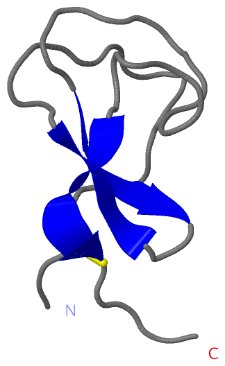
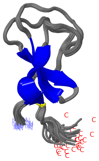
NMR Structure
|
||||||||
Cis Peptide Bonds (0, 0)| (no "Cis Peptide Bond" information available for 2D49) |
SAPs(SNPs)/Variants (0, 0)| (no "SAP(SNP)/Variant" information available for 2D49) |
PROSITE Motifs (0, 0)| (no "PROSITE Motif" information available for 2D49) |
Exons (0, 0)| (no "Exon" information available for 2D49) |
Sequences/Alignments
NMR StructureChain A from PDB Type:PROTEIN Length:53 aligned with O50152_STRGR | O50152 from UniProtKB/TrEMBL Length:294 Alignment length:53 39 49 59 69 79 O50152_STRGR 30 ATCATAWSSSSVYTNGGTVSYNGRNYTAKWWTQNERPGTSDVWADKGACGTGG 82 SCOP domains ----------------------------------------------------- SCOP domains CATH domains ----------------------------------------------------- CATH domains Pfam domains ----------------------------------------------------- Pfam domains SAPs(SNPs) ----------------------------------------------------- SAPs(SNPs) PROSITE ----------------------------------------------------- PROSITE Transcript ----------------------------------------------------- Transcript 2d49 A 1 ATCATAWSSSSVYTNGGTVSYNGRNYTAKWWTQNERPGTSDVWADKGACGTGS 53 10 20 30 40 50
|
||||||||||||||||||||
SCOP Domains (0, 0)| (no "SCOP Domain" information available for 2D49) |
CATH Domains (0, 0)| (no "CATH Domain" information available for 2D49) |
Pfam Domains (0, 0)| (no "Pfam Domain" information available for 2D49) |
Gene Ontology (7, 7)|
NMR Structure(hide GO term definitions) Chain A (O50152_STRGR | O50152)
|
||||||||||||||||||||||||||||||||||||||||||||||||||||||||||||
Interactive Views
|
||||||||||||||||||||||||||||||||||||||||||||||||||||||||||||||||||||||||||||||||||||||||||||||||||||||||||||||||||||
Still Images
|
||||||||||||||||
Databases
|
||||||||||||||||||||||||||||||||||||||||||||||||||||||||||||||||||||||||||||||||||||||||||||||||||||||||||||||||||||||||||||||||||||||||||||||||||||||||||||||||
Analysis Tools
|
|||||||||||||||||||||||||||||||||||||||||||||||||||||||||||||
Entries Sharing at Least One Protein Chain (UniProt ID)
Related Entries Specified in the PDB File
|
|