Show PDB file:   
         Plain Text   HTML   (compressed file size)
QuickSearch:   
by PDB,NDB,UniProt,PROSITE Code or Search Term(s)  
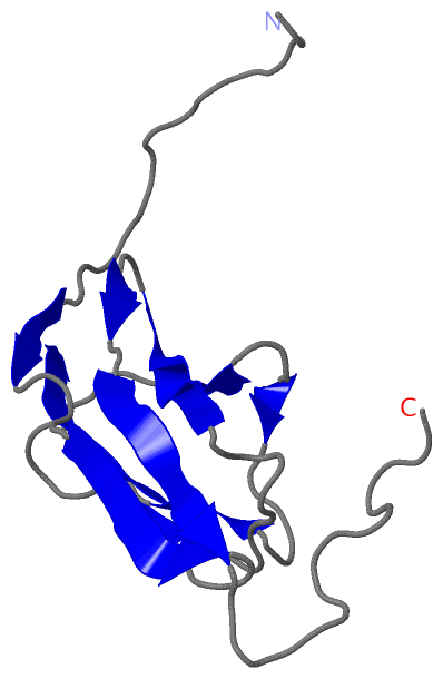
(-)NMR Structure - model 1
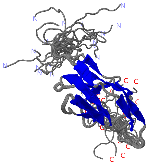
(-)NMR Structure - all models
collapse expand < >
Image NMR Structure - model 1
NMR Structure - model 1  (Jmol Viewer)
Image NMR Structure - all models
NMR Structure - all models  (Jmol Viewer)

(-) Description

Title :  SOLUTION STRUCTURE OF THE IG DOMAIN (2998-3100) OF HUMAN OBSCURIN
 
Authors :  K. Nagashima, F. Hayashi, M. Yoshida, S. Yokoyama, Riken Structural Genomics/Proteomics Initiative (Rsgi)
Date :  20 May 05  (Deposition) - 20 Nov 05  (Release) - 24 Feb 09  (Revision)
Method :  SOLUTION NMR
Resolution :  NOT APPLICABLE
Chains :  NMR Structure  :  A  (20x)
Keywords :  Ig-Fold, Kiaa1556 Protein, Immunoglobulin Domain, Structural Genomics, Nppsfa, National Project On Protein Structural And Functional Analyses, Riken Structural Genomics/Proteomics Initiative, Rsgi, Contractile Protein (Keyword Search: [Gene Ontology, PubMed, Web (Google)] )
 
Reference :  K. Nagashima, F. Hayashi, M. Yoshida, S. Yokoyama
Solution Structure Of The Ig Domain (2998-3100) Of Human Obscurin
To Be Published
PubMed: search
(for further references see the PDB file header)

(-) Compounds

Molecule 1 - KIAA1556 PROTEIN
    Chains: A
    Engineered: YES
    Expression System Plasmid: P040202-82
    Expression System Vector Type: PLASMID
    Fragment: IG
    Gene: KIAA1556
    Organism Common: HUMAN
    Organism Scientific: HOMO SAPIENS
    Organism Taxid: 9606
    Other Details: CELL FREE PROTEIN SYNTHESIS
    Synonym: OBSCURIN

 Structural Features

(-) Chains, Units

  
NMR Structure (20x): 

Summary Information (see also Sequences/Alignments below)

(-) Ligands, Modified Residues, Ions  (0, 0)

(no "Ligand,Modified Residues,Ions" information available for 2CR6)

(-) Sites  (0, 0)

(no "Site" information available for 2CR6)

(-) SS Bonds  (0, 0)

(no "SS Bond" information available for 2CR6)

(-) Cis Peptide Bonds  (2, 40)

NMR Structure
No.ModelResidues
11, 2, 3, 4, 5, 6, 7, 8, 9, 10, 11, 12, 13, 14, 15, 16, 17, 18, 19, 20Ser A:38 -Pro A:39
21, 2, 3, 4, 5, 6, 7, 8, 9, 10, 11, 12, 13, 14, 15, 16, 17, 18, 19, 20Glu A:43 -Pro A:44

 Sequence-Structure Mapping

(-) SAPs(SNPs)/Variants  (0, 0)

(no "SAP(SNP)/Variant" information available for 2CR6)

(-) PROSITE Motifs  (0, 0)

(no "PROSITE Motif" information available for 2CR6)

(-) Exons   (0, 0)

(no "Exon" information available for 2CR6)

(-) Sequences/Alignments

NMR Structure
   Reformat: Number of residues per line =  ('0' or empty: single-line sequence representation)
  Number of residues per labelling interval =   
  UniProt sequence: complete  aligned part    
   Show mapping: SCOP domains CATH domains Pfam domains Secondary structure (by author)
SAPs(SNPs) PROSITE motifs Exons
(details for a mapped element are shown in a popup box when the mouse pointer rests over it)
Chain A from PDB  Type:PROTEIN  Length:115
 aligned with OBSCN_HUMAN | Q5VST9 from UniProtKB/Swiss-Prot  Length:7968

    Alignment length:174
                                  2999      3009      3019      3029      3039      3049      3059      3069      3079      3089      3099      3109      3119      3129      3139      3149      3159    
         OBSCN_HUMAN   2990 GQAQSRAQLLVQGRRVHIIEDLEDVDVQEGSSATFRCRISPANYEPVHWFLDKTPLHANELNEIDAQPGGYHVLTLRQLALKDSGTIYFEAGDQRASAALRVTEKPSVFSRELTDATITEGEDLTLVCETSTCDIPVCWTKDGKTLRGSARCQLSHEGHRAQLLITGATLQDSG 3163
               SCOP domains ------------------------------------------------------------------------------------------------------------------------------------------------------------------------------ SCOP domains
               CATH domains ------------------------------------------------------------------------------------------------------------------------------------------------------------------------------ CATH domains
               Pfam domains ------------------------------------------------------------------------------------------------------------------------------------------------------------------------------ Pfam domains
         Sec.struct. author .......--.......eee....eeeee....eeeeeeee.......eeee...ee.....eee......eeeeee........eeeeeee..ee..eeeeee.........-----------------------------------...----------------------.. Sec.struct. author
                 SAPs(SNPs) ------------------------------------------------------------------------------------------------------------------------------------------------------------------------------ SAPs(SNPs)
                    PROSITE ------------------------------------------------------------------------------------------------------------------------------------------------------------------------------ PROSITE
                 Transcript ------------------------------------------------------------------------------------------------------------------------------------------------------------------------------ Transcript
                2cr6 A    1 GSSGSSG--LVQGRRVHIIEDLEDVDVQEGSSATFRCRISPANYEPVHWFLDKTPLHANELNEIDAQPGGYHVLTLRQLALKDSGTIYFEAGDQRASAALRVTEKPSVFSRS-----------------------------------GPS----------------------SG  115
                                  |  8        18        28        38        48        58        68        78        88        98       108 |       -         -         -       113         -         -  | 
                                  7  8                                                                                                   110                                 111 |                    114 
                                                                                                                                                                               113                        

   Legend:   → Mismatch (orange background)
  - → Gap (green background, '-', border residues have a numbering label)
    → Modified Residue (blue background, lower-case, 'x' indicates undefined single-letter code, labelled with number + name)
  x → Chemical Group (purple background, 'x', labelled with number + name, e.g. ACE or NH2)
  extra numbering lines below/above indicate numbering irregularities and modified residue names etc., number ends below/above '|'

 Classification and Annotation

(-) SCOP Domains  (0, 0)

(no "SCOP Domain" information available for 2CR6)

(-) CATH Domains  (0, 0)

(no "CATH Domain" information available for 2CR6)

(-) Pfam Domains  (0, 0)

(no "Pfam Domain" information available for 2CR6)

(-) Gene Ontology  (30, 30)

NMR Structure(hide GO term definitions)
Chain A   (OBSCN_HUMAN | Q5VST9)
molecular function
    GO:0005524    ATP binding    Interacting selectively and non-covalently with ATP, adenosine 5'-triphosphate, a universally important coenzyme and enzyme regulator.
    GO:0005089    Rho guanyl-nucleotide exchange factor activity    Stimulates the exchange of guanyl nucleotides associated with a GTPase of the Rho family. Under normal cellular physiological conditions, the concentration of GTP is higher than that of GDP, favoring the replacement of GDP by GTP in association with the GTPase.
    GO:0030506    ankyrin binding    Interacting selectively and non-covalently with ankyrin, a 200 kDa cytoskeletal protein that attaches other cytoskeletal proteins to integral membrane proteins.
    GO:0005516    calmodulin binding    Interacting selectively and non-covalently with calmodulin, a calcium-binding protein with many roles, both in the calcium-bound and calcium-free states.
    GO:0005085    guanyl-nucleotide exchange factor activity    Stimulates the exchange of guanyl nucleotides associated with a GTPase. Under normal cellular physiological conditions, the concentration of GTP is higher than that of GDP, favoring the replacement of GDP by GTP in association with the GTPase.
    GO:0016301    kinase activity    Catalysis of the transfer of a phosphate group, usually from ATP, to a substrate molecule.
    GO:0046872    metal ion binding    Interacting selectively and non-covalently with any metal ion.
    GO:0000166    nucleotide binding    Interacting selectively and non-covalently with a nucleotide, any compound consisting of a nucleoside that is esterified with (ortho)phosphate or an oligophosphate at any hydroxyl group on the ribose or deoxyribose.
    GO:0005515    protein binding    Interacting selectively and non-covalently with any protein or protein complex (a complex of two or more proteins that may include other nonprotein molecules).
    GO:0004672    protein kinase activity    Catalysis of the phosphorylation of an amino acid residue in a protein, usually according to the reaction: a protein + ATP = a phosphoprotein + ADP.
    GO:0004674    protein serine/threonine kinase activity    Catalysis of the reactions: ATP + protein serine = ADP + protein serine phosphate, and ATP + protein threonine = ADP + protein threonine phosphate.
    GO:0008307    structural constituent of muscle    The action of a molecule that contributes to the structural integrity of a muscle fiber.
    GO:0031432    titin binding    Interacting selectively and non-covalently with titin, any of a family of giant proteins found in striated and smooth muscle. In striated muscle, single titin molecules span half the sarcomere, with their N- and C-termini in the Z-disc and M-line, respectively.
    GO:0016740    transferase activity    Catalysis of the transfer of a group, e.g. a methyl group, glycosyl group, acyl group, phosphorus-containing, or other groups, from one compound (generally regarded as the donor) to another compound (generally regarded as the acceptor). Transferase is the systematic name for any enzyme of EC class 2.
biological process
    GO:0030154    cell differentiation    The process in which relatively unspecialized cells, e.g. embryonic or regenerative cells, acquire specialized structural and/or functional features that characterize the cells, tissues, or organs of the mature organism or some other relatively stable phase of the organism's life history. Differentiation includes the processes involved in commitment of a cell to a specific fate and its subsequent development to the mature state.
    GO:0007275    multicellular organism development    The biological process whose specific outcome is the progression of a multicellular organism over time from an initial condition (e.g. a zygote or a young adult) to a later condition (e.g. a multicellular animal or an aged adult).
    GO:0016310    phosphorylation    The process of introducing a phosphate group into a molecule, usually with the formation of a phosphoric ester, a phosphoric anhydride or a phosphoric amide.
    GO:0043547    positive regulation of GTPase activity    Any process that activates or increases the activity of a GTPase.
    GO:0043065    positive regulation of apoptotic process    Any process that activates or increases the frequency, rate or extent of cell death by apoptotic process.
    GO:0098779    positive regulation of macromitophagy in response to mitochondrial depolarization    The macromitophagy process that is triggered by a detection of the loss of mitochondrial membrane potential.
    GO:0036309    protein localization to M-band    Any process in which a protein is transported to, and/or maintained in, the M band. The M band is the midline of aligned thick filaments in a sarcomere.
    GO:0006468    protein phosphorylation    The process of introducing a phosphate group on to a protein.
    GO:0035023    regulation of Rho protein signal transduction    Any process that modulates the frequency, rate or extent of Rho protein signal transduction.
    GO:0051056    regulation of small GTPase mediated signal transduction    Any process that modulates the frequency, rate or extent of small GTPase mediated signal transduction.
    GO:0045214    sarcomere organization    The myofibril assembly process that results in the organization of muscle actomyosin into sarcomeres. The sarcomere is the repeating unit of a myofibril in a muscle cell, composed of an array of overlapping thick and thin filaments between two adjacent Z discs.
cellular component
    GO:0031430    M band    The midline of aligned thick filaments in a sarcomere; location of specific proteins that link thick filaments. Depending on muscle type the M band consists of different numbers of M lines.
    GO:0030018    Z disc    Platelike region of a muscle sarcomere to which the plus ends of actin filaments are attached.
    GO:0005737    cytoplasm    All of the contents of a cell excluding the plasma membrane and nucleus, but including other subcellular structures.
    GO:0005829    cytosol    The part of the cytoplasm that does not contain organelles but which does contain other particulate matter, such as protein complexes.
    GO:0030016    myofibril    The contractile element of skeletal and cardiac muscle; a long, highly organized bundle of actin, myosin, and other proteins that contracts by a sliding filament mechanism.

 Visualization

(-) Interactive Views

NMR Structure
  Complete Structure
    Jena3D(integrated viewing of ligand, site, SAP, PROSITE, SCOP information)
    WebMol | AstexViewer[tm]@PDBe
(Java Applets, require no local installation except for Java; loading may be slow)
    STRAP
(Java WebStart application, automatic local installation, requires Java; full application with system access!)
    RasMol
(require local installation)
    Molscript (VRML)
(requires installation of a VRML viewer; select preferred view via VRML and generate a mono or stereo PDF format file)
 
  Ligands, Modified Residues, Ions
(no "Ligands, Modified Residues, Ions" information available for 2cr6)
 
  Sites
(no "Sites" information available for 2cr6)
 
  Cis Peptide Bonds
    Glu A:43 - Pro A:44   [ RasMol ]  
    Ser A:38 - Pro A:39   [ RasMol ]  
 

(-) Still Images

Jmol
  protein: cartoon or spacefill or dots and stick; nucleic acid: cartoon and stick; ligands: spacefill; active site: stick
Molscript
  protein, nucleic acid: cartoon; ligands: spacefill; active site: ball and stick

 Databases and Analysis Tools

(-) Databases

Access by PDB/NDB ID
  2cr6
    Family and Domain Information: ProDom | SYSTERS
    General Structural Information: GlycoscienceDB | MMDB | NDB | OCA | PDB | PDBe | PDBj | PDBsum | PDBWiki | PQS | PROTEOPEDIA
    Orientation in Membranes: OPM
    Protein Surface: SURFACE
    Secondary Structure: DSSP (structure derived) | HSSP (homology derived)
    Structural Genomics: GeneCensus
    Structural Neighbours: CE | VAST
    Structure Classification: CATH | Dali | SCOP
    Validation and Original Data: BMRB Data View | BMRB Restraints Grid | EDS | PROCHECK | RECOORD | WHAT_CHECK
 
Access by UniProt ID/Accession number
  OBSCN_HUMAN | Q5VST9
    Comparative Protein Structure Models: ModBase
    Genomic Information: Ensembl
    Protein-protein Interaction: DIP
    Sequence, Family and Domain Information: InterPro | Pfam | SMART | UniProtKB/SwissProt
 
Access by Enzyme Classificator   (EC Number)
  (no 'Enzyme Classificator' available)
    General Enzyme Information: BRENDA | EC-PDB | Enzyme | IntEnz
    Pathway: KEGG | MetaCyc
 
Access by Disease Identifier   (MIM ID)
  (no 'MIM ID' available)
    Disease Information: OMIM
 
Access by GenAge ID
  (no 'GenAge ID' available)
    Age Related Information: GenAge

(-) Analysis Tools

Access by PDB/NDB ID
    Domain Information: XDom
    Interatomic Contacts of Structural Units: CSU
    Ligand-protein Contacts: LPC
    Protein Cavities: castP
    Sequence and Secondary Structure: PDBCartoon
    Structure Alignment: STRAP(Java WebStart application, automatic local installation, requires Java; full application with system access!)
    Structure and Sequence Browser: STING
 
Access by UniProt ID/Accession number
  OBSCN_HUMAN | Q5VST9
    Protein Disorder Prediction: DisEMBL | FoldIndex | GLOBPLOT (for more information see DisProt)

 Related Entries

(-) Entries Sharing at Least One Protein Chain (UniProt ID)

UniProtKB/Swiss-Prot
        OBSCN_HUMAN | Q5VST9: 1v1c 2dku 2dm7 2e7b 2edf 2edh 2edl 2edq 2edr 2edt 2edw 2eny 2eo1 2gqh 2mwc 2n56 2yz8 4c4k 4rsv 4uow

(-) Related Entries Specified in the PDB File

(no "Related Entries Specified in the PDB File" available for 2CR6)