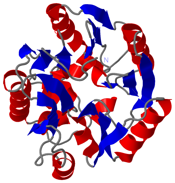
Show PDB file:   
         Plain Text   HTML   (compressed file size)
QuickSearch:   
by PDB,NDB,UniProt,PROSITE Code or Search Term(s)  
(-)Asym./Biol. Unit
collapse expand < >
Image Asym./Biol. Unit
Asym./Biol. Unit  (Jmol Viewer)

(-) Description

Title :  CRYSTAL STRUCTURE OF DIMETHYLARGININE DIMETHYLAMINOHYDROLASE CRYSTAL FORM I
 
Authors :  D. Frey, O. Braun, C. Briand, M. Vasak, M. G. Grutter
Date :  17 Mar 06  (Deposition) - 17 May 06  (Release) - 24 Feb 09  (Revision)
Method :  X-RAY DIFFRACTION
Resolution :  1.70
Chains :  Asym./Biol. Unit :  A
Keywords :  Nos Regulation, S-Nitrosylation, Zinc, Hydrolase, No, Mma, Adma (Keyword Search: [Gene Ontology, PubMed, Web (Google))
 
Reference :  D. Frey, O. Braun, C. Briand, M. Vasak, M. G. Grutter
Structure Of The Mammalian Nos Regulator Dimethylarginine Dimethylaminohydrolase: A Basis For The Design Of Specific Inhibitors.
Structure V. 14 901 2006
PubMed-ID: 16698551  |  Reference-DOI: 10.1016/J.STR.2006.03.006

(-) Compounds

Molecule 1 - NG, NG-DIMETHYLARGININE DIMETHYLAMINOHYDROLASE 1
    ChainsA
    EC Number3.5.3.18
    OrganBRAIN
    Organism CommonBOVINE
    Organism ScientificBOS TAURUS
    Organism Taxid9913
    SynonymDIMETHYLARGININE DIMETHYLAMINOHYDROLASE I, DIMETHYLARGININASE 1, DDAHI, DDAH-1

 Structural Features

(-) Chains, Units

  1
Asymmetric/Biological Unit A

Summary Information (see also Sequences/Alignments below)

(-) Ligands, Modified Residues, Ions  (0, 0)

(no "Ligand,Modified Residues,Ions" information available for 2CI3)

(-) Sites  (0, 0)

(no "Site" information available for 2CI3)

(-) SS Bonds  (0, 0)

(no "SS Bond" information available for 2CI3)

(-) Cis Peptide Bonds  (0, 0)

(no "Cis Peptide Bond" information available for 2CI3)

 Sequence-Structure Mapping

(-) SAPs(SNPs)/Variants  (0, 0)

(no "SAP(SNP)/Variant" information available for 2CI3)

(-) PROSITE Motifs  (0, 0)

(no "PROSITE Motif" information available for 2CI3)

(-) Exons   (0, 0)

(no "Exon" information available for 2CI3)

(-) Sequences/Alignments

Asymmetric/Biological Unit
   Reformat: Number of residues per line =  ('0' or empty: single-line sequence representation)
  Number of residues per labelling interval =   
  UniProt sequence: complete  aligned part    
   Show mapping: SCOP domains CATH domains Pfam domains Secondary structure (by author)
SAPs(SNPs) PROSITE motifs Exons
(details for a mapped element are shown in a popup box when the mouse pointer rests over it)
Chain A from PDB  Type:PROTEIN  Length:274
 aligned with DDAH1_BOVIN | P56965 from UniProtKB/Swiss-Prot  Length:285

    Alignment length:274
                                    17        27        37        47        57        67        77        87        97       107       117       127       137       147       157       167       177       187       197       207       217       227       237       247       257       267       277    
          DDAH1_BOVIN     8 ATFGRATHVVVRALPESLAQQALRRTKGDEVDFARAERQHQLYVGVLGSKLGLQVVQLPADESLPDCVFVEDVAVVCEETALITRPGAPSRRKEADMMKEALEKLQLNIVEMKDENATLDGGDVLFTGREFFVGLSKRTNQRGAEILADTFKDYAVSTVPVVDALHLKSFCSMAGPNLIAIGSSESAQKALKIMQQMSDHRYDKLTVPDDTAANCIYLNIPSKGHVLLHRTPEEYPESAKVYEKLKDHMLIPVSNSELEKVDGLLTCSSVLINK 281
               SCOP domains d2ci3a_ A: automated matches                                                                                                                                                                                                                                                       SCOP domains
               CATH domains ---------------------------------------------------------------------------------------------------------------------------------------------------------------------------------------------------------------------------------------------------------------------------------- CATH domains
               Pfam domains ---------------------------------------------------------------------------------------------------------------------------------------------------------------------------------------------------------------------------------------------------------------------------------- Pfam domains
         Sec.struct. author .......eeeee....hhhhhh.........hhhhhhhhhhhhhhhhhh....eeeee..........hhhh.eeee..eeee....hhhhhhhhhhhhhhhhhh..eeee........hhh.eee...eeeeee....hhhhhhhhhhhh...eeeeee.....hhhh.eeeee..eeeee.hhhhhhhhhhhhhh.....eeeee.hhhhhh.eeeee...eeeeee.....hhhhhhhhh.....eeee..hhhhhh...hhhhhheee.. Sec.struct. author
                 SAPs(SNPs) ---------------------------------------------------------------------------------------------------------------------------------------------------------------------------------------------------------------------------------------------------------------------------------- SAPs(SNPs)
                    PROSITE ---------------------------------------------------------------------------------------------------------------------------------------------------------------------------------------------------------------------------------------------------------------------------------- PROSITE
                 Transcript ---------------------------------------------------------------------------------------------------------------------------------------------------------------------------------------------------------------------------------------------------------------------------------- Transcript
                 2ci3 A   7 ATFGRATHVVVRALPESLAQQALRRTKGDEVDFARAERQHQLYVGVLGSKLGLQVVQLPADESLPDCVFVEDVAVVCEETALITRPGAPSRRKEADMMKEALEKLQLNIVEMKDENATLDGGDVLFTGREFFVGLSKRTNQRGAEILADTFKDYAVSTVPVVDALHLKSFCSMAGPNLIAIGSSESAQKALKIMQQMSDHRYDKLTVPDDTAANCIYLNIPSKGHVLLHRTPEEYPESAKVYEKLKDHMLIPVSNSELEKVDGLLTCSSVLINK 280
                                    16        26        36        46        56        66        76        86        96       106       116       126       136       146       156       166       176       186       196       206       216       226       236       246       256       266       276    

   Legend:   → Mismatch (orange background)
  - → Gap (green background, '-', border residues have a numbering label)
    → Modified Residue (blue background, lower-case, 'x' indicates undefined single-letter code, labelled with number + name)
  x → Chemical Group (purple background, 'x', labelled with number + name, e.g. ACE or NH2)
  extra numbering lines below/above indicate numbering irregularities and modified residue names etc., number ends below/above '|'

 Classification and Annotation

(-) SCOP Domains  (1, 1)

Asymmetric/Biological Unit

(-) CATH Domains  (0, 0)

(no "CATH Domain" information available for 2CI3)

(-) Pfam Domains  (0, 0)

(no "Pfam Domain" information available for 2CI3)

(-) Gene Ontology  (12, 12)

Asymmetric/Biological Unit(hide GO term definitions)
Chain A   (DDAH1_BOVIN | P56965)
molecular function
    GO:0016403    dimethylargininase activity    Catalysis of the reaction: N(G),N(G)-dimethyl-L-arginine + H2O = dimethylamine + L-citrulline.
    GO:0016787    hydrolase activity    Catalysis of the hydrolysis of various bonds, e.g. C-O, C-N, C-C, phosphoric anhydride bonds, etc. Hydrolase is the systematic name for any enzyme of EC class 3.
    GO:0046872    metal ion binding    Interacting selectively and non-covalently with any metal ion.
    GO:0008270    zinc ion binding    Interacting selectively and non-covalently with zinc (Zn) ions.
biological process
    GO:0006527    arginine catabolic process    The chemical reactions and pathways resulting in the breakdown of arginine, 2-amino-5-(carbamimidamido)pentanoic acid.
    GO:0000052    citrulline metabolic process    The chemical reactions and pathways involving citrulline, N5-carbamoyl-L-ornithine, an alpha amino acid not found in proteins.
    GO:0006809    nitric oxide biosynthetic process    The chemical reactions and pathways resulting in the formation of nitric oxide, nitrogen monoxide (NO), a colorless gas only slightly soluble in water.
    GO:0045429    positive regulation of nitric oxide biosynthetic process    Any process that activates or increases the frequency, rate or extent of the chemical reactions and pathways resulting in the formation of nitric oxide.
    GO:0017014    protein nitrosylation    The covalent addition of a nitric oxide group to an amino acid within a protein.
    GO:0003073    regulation of systemic arterial blood pressure    The process that modulates the force with which blood travels through the systemic arterial circulatory system. The process is controlled by a balance of processes that increase pressure and decrease pressure.
cellular component
    GO:0070062    extracellular exosome    A vesicle that is released into the extracellular region by fusion of the limiting endosomal membrane of a multivesicular body with the plasma membrane. Extracellular exosomes, also simply called exosomes, have a diameter of about 40-100 nm.
    GO:0005739    mitochondrion    A semiautonomous, self replicating organelle that occurs in varying numbers, shapes, and sizes in the cytoplasm of virtually all eukaryotic cells. It is notably the site of tissue respiration.

 Visualization

(-) Interactive Views

Asymmetric/Biological Unit
  Complete Structure
    Jena3D(integrated viewing of ligand, site, SAP, PROSITE, SCOP information)
    WebMol | AstexViewer[tm]@PDBe
(Java Applets, require no local installation except for Java; loading may be slow)
    STRAP
(Java WebStart application, automatic local installation, requires Java; full application with system access!)
    RasMol
(require local installation)
    Molscript (VRML)
(requires installation of a VRML viewer; select preferred view via VRML and generate a mono or stereo PDF format file)
 
  Ligands, Modified Residues, Ions
(no "Ligands, Modified Residues, Ions" information available for 2ci3)
 
  Sites
(no "Sites" information available for 2ci3)
 
  Cis Peptide Bonds
(no "Cis Peptide Bonds" information available for 2ci3)
 

(-) Still Images

Jmol
  protein: cartoon or spacefill or dots and stick; nucleic acid: cartoon and stick; ligands: spacefill; active site: stick
Molscript
  protein, nucleic acid: cartoon; ligands: spacefill; active site: ball and stick

 Databases and Analysis Tools

(-) Databases

Access by PDB/NDB ID
  2ci3
    Family and Domain InformationProDom | SYSTERS
    General Structural InformationGlycoscienceDB | MMDB | NDB | OCA | PDB | PDBe | PDBj | PDBsum | PDBWiki | PQS | PROTEOPEDIA
    Orientation in MembranesOPM
    Protein SurfaceSURFACE
    Secondary StructureDSSP (structure derived) | HSSP (homology derived)
    Structural GenomicsGeneCensus
    Structural NeighboursCE | VAST
    Structure ClassificationCATH | Dali | SCOP
    Validation and Original DataBMRB Data View | BMRB Restraints Grid | EDS | PROCHECK | RECOORD | WHAT_CHECK
 
Access by UniProt ID/Accession number
  DDAH1_BOVIN | P56965
    Comparative Protein Structure ModelsModBase
    Genomic InformationEnsembl
    Protein-protein InteractionDIP
    Sequence, Family and Domain InformationInterPro | Pfam | SMART | UniProtKB/SwissProt
 
Access by Enzyme Classificator   (EC Number)
  3.5.3.18
    General Enzyme InformationBRENDA | EC-PDB | Enzyme | IntEnz
    PathwayKEGG | MetaCyc
 
Access by Disease Identifier   (MIM ID)
  (no 'MIM ID' available)
    Disease InformationOMIM
 
Access by GenAge ID
  (no 'GenAge ID' available)
    Age Related InformationGenAge

(-) Analysis Tools

Access by PDB/NDB ID
    Domain InformationXDom
    Interatomic Contacts of Structural UnitsCSU
    Ligand-protein ContactsLPC
    Protein CavitiescastP
    Sequence and Secondary StructurePDBCartoon
    Structure AlignmentSTRAP(Java WebStart application, automatic local installation, requires Java; full application with system access!)
    Structure and Sequence BrowserSTING
 
Access by UniProt ID/Accession number
  DDAH1_BOVIN | P56965
    Protein Disorder PredictionDisEMBL | FoldIndex | GLOBPLOT (for more information see DisProt)

 Related Entries

(-) Entries Sharing at Least One Protein Chain (UniProt ID)

UniProtKB/Swiss-Prot
        DDAH1_BOVIN | P569652c6z 2ci1 2ci4 2ci5 2ci6 2ci7

(-) Related Entries Specified in the PDB File

2c6z CRYSTAL STRUCTURE OF DIMETHYLARGININE DIMETHYLAMINOHYDROLASE I IN COMPLEX WITH CITRULLINE
2ci1 CRYSTAL STRUCTURE OF DIMETHYLARGININE DIMETHYLAMINOHYDROLASE I IN COMPLEX WITH S- NITROSO-LHOMOCYSTEINE
2ci4 CRYSTAL STRUCTURE OF DIMETHYLARGININE DIMETHYLAMINOHYDROLASE I CRYSTAL FORM II
2ci5 CRYSTAL STRUCTURE OF DIMETHYLARGININE DIMETHYLAMINOHYDROLASE I IN COMPLEX WITH L- HOMOCYSTEINE
2ci6 CRYSTAL STRUCTURE OF DIMETHYLARGININE DIMETHYLAMINOHYDROLASE I BOUND WITH ZINC LOW PH
2ci7 CRYSTAL STRUCTURE OF DIMETHYLARGININE DIMETHYLAMINOHYDROLASE I IN COMPLEX WITH ZINC , HIGH PH