Show PDB file:   
         Plain Text   HTML   (compressed file size)
QuickSearch:   
by PDB,NDB,UniProt,PROSITE Code or Search Term(s)  
(-)Asym./Biol. Unit
(-)Asym./Biol. Unit - sites
collapse expand < >
Image Asym./Biol. Unit
Asym./Biol. Unit  (Jmol Viewer)
Image Asym./Biol. Unit - sites
Asym./Biol. Unit - sites  (Jmol Viewer)

(-) Description

Title :  CELLULAR RETINOIC ACID BINDING PROTEIN I IN COMPLEX WITH A RETINOBENZOIC ACID (AM80)
 
Authors :  B. Chaudhuri, G. J. Kleywegt, T. Bergfors, T. A. Jones
Date :  22 Feb 99  (Deposition) - 21 Dec 99  (Release) - 05 Oct 11  (Revision)
Method :  X-RAY DIFFRACTION
Resolution :  2.80
Chains :  Asym./Biol. Unit :  A
Keywords :  Retinoic-Acid Transport, Transport Protein (Keyword Search: [Gene Ontology, PubMed, Web (Google))
 
Reference :  B. N. Chaudhuri, G. J. Kleywegt, I. Broutin-L'Hermite, T. Bergfors, H. Senn, P. Le Motte, O. Partouche, T. A. Jones
Structures Of Cellular Retinoic Acid Binding Proteins I And Ii In Complex With Synthetic Retinoids.
Acta Crystallogr. , Sect. D V. 55 1850 1999
PubMed-ID: 10531482  |  Reference-DOI: 10.1107/S0907444999011026
(for further references see the PDB file header)

(-) Compounds

Molecule 1 - PROTEIN (CRABP-I)
    ChainsA
    EngineeredYES
    Expression SystemESCHERICHIA COLI BL21(DE3)
    Expression System Cellular LocationCYTOPLASM
    Expression System PlasmidPT7-1-3
    Expression System StrainBL21(DE3)
    Expression System Taxid469008
    GeneMOUSE
    Organism CommonCATTLE
    Organism ScientificBOS TAURUS
    Organism Taxid9913
    Other DetailsMUS MUSCULUS (IDENTICAL WITH BOVINE PROTEIN) RECOMBINANT GENE

 Structural Features

(-) Chains, Units

  1
Asymmetric/Biological Unit A

Summary Information (see also Sequences/Alignments below)

(-) Ligands, Modified Residues, Ions  (1, 1)

Asymmetric/Biological Unit (1, 1)
No.NameCountTypeFull Name
1A801Ligand/Ion4-[(5,5,8,8-TETRAMETHYL-5,6,7,8-TETRAHYDRONAPHTHALEN-2-YL)CARBAMOYL]BENZOIC ACID

(-) Sites  (1, 1)

Asymmetric Unit (1, 1)
No.NameEvidenceResiduesDescription
1AC1SOFTWARETHR A:54 , THR A:56 , VAL A:58 , ARG A:59 , VAL A:76 , ASP A:77 , GLY A:78 , LEU A:120 , ARG A:131 , TYR A:133 , HOH A:303 , HOH A:306BINDING SITE FOR RESIDUE A80 A 201

(-) SS Bonds  (0, 0)

(no "SS Bond" information available for 2CBR)

(-) Cis Peptide Bonds  (0, 0)

(no "Cis Peptide Bond" information available for 2CBR)

 Sequence-Structure Mapping

(-) SAPs(SNPs)/Variants  (0, 0)

(no "SAP(SNP)/Variant" information available for 2CBR)

(-) PROSITE Motifs  (1, 1)

Asymmetric/Biological Unit (1, 1)
 PROSITEUniProtKBPDB
No.IDACDescriptionIDLocationCountLocation
1FABPPS00214 Cytosolic fatty-acid binding proteins signature.RABP1_BOVIN6-23  1A:5-22

(-) Exons   (4, 4)

Asymmetric/Biological Unit (4, 4)
 ENSEMBLUniProtKBPDB
No.Transcript IDExonExon IDGenome LocationLengthIDLocationLengthCountLocationLength
1.1ENSBTAT000000171581ENSBTAE00000139322chr21:30790357-30790713357RABP1_BOVIN1-32321A:1-3131
1.2ENSBTAT000000171582ENSBTAE00000139323chr21:30791249-30791433185RABP1_BOVIN32-93621A:31-9262
1.3ENSBTAT000000171583ENSBTAE00000139324chr21:30793481-30793594114RABP1_BOVIN94-131381A:93-13038
1.4ENSBTAT000000171584ENSBTAE00000139325chr21:30797275-30797552278RABP1_BOVIN132-147161A:131-1366

(-) Sequences/Alignments

Asymmetric/Biological Unit
   Reformat: Number of residues per line =  ('0' or empty: single-line sequence representation)
  Number of residues per labelling interval =   
  UniProt sequence: complete  aligned part    
   Show mapping: SCOP domains CATH domains Pfam domains Secondary structure (by author)
SAPs(SNPs) PROSITE motifs Exons
(details for a mapped element are shown in a popup box when the mouse pointer rests over it)
Chain A from PDB  Type:PROTEIN  Length:136
 aligned with RABP1_BOVIN | P62964 from UniProtKB/Swiss-Prot  Length:137

    Alignment length:136
                                    11        21        31        41        51        61        71        81        91       101       111       121       131      
          RABP1_BOVIN     2 PNFAGTWKMRSSENFDELLKALGVNAMLRKVAVAAASKPHVEIRQDGDQFYIKTSTTVRTTEINFKVGEGFEEETVDGRKCRSLPTWENENKIHCTQTLLEGDGPKTYWTRELANDELILTFGADDVVCTRIYVRE 137
               SCOP domains d2cbra_ A: Cellular retinoic-acid-binding protein (CRABP)                                                                                SCOP domains
               CATH domains 2cbrA00 A:1-136  [code=2.40.128.20, no name defined]                                                                                     CATH domains
               Pfam domains ---------------------------------------------------------------------------------------------------------------------------------------- Pfam domains
         Sec.struct. author ....eeeeeeeee.hhhhhhh....hhhhhhhhhhhhh.eeeeeee..eeeeeee....eeeeeee....eeee.....eeeeeeeeee..eeeeeeee.......eeeeeeee..eeeeeeee..eeeeeeeee. Sec.struct. author
                 SAPs(SNPs) ---------------------------------------------------------------------------------------------------------------------------------------- SAPs(SNPs)
                    PROSITE ----FABP  PDB: A:5-22 ------------------------------------------------------------------------------------------------------------------ PROSITE
           Transcript 1 (1) Exon 1.1  PDB: A:1-31          -------------------------------------------------------------Exon 1.3  PDB: A:93-130               1.4    Transcript 1 (1)
           Transcript 1 (2) ------------------------------Exon 1.2  PDB: A:31-92 UniProt: 32-93                         -------------------------------------------- Transcript 1 (2)
                 2cbr A   1 PNFAGTWKMRSSENFDELLKALGVNAMLRKVAVAAASKPHVEIRQDGDQFYIKTSTTVRTTEINFKVGEGFEEETVDGRKCRSLPTWENENKIHCTQTLLEGDGPKTYWTRELANDELILTFGADDVVCTRIYVRE 136
                                    10        20        30        40        50        60        70        80        90       100       110       120       130      

   Legend:   → Mismatch (orange background)
  - → Gap (green background, '-', border residues have a numbering label)
    → Modified Residue (blue background, lower-case, 'x' indicates undefined single-letter code, labelled with number + name)
  x → Chemical Group (purple background, 'x', labelled with number + name, e.g. ACE or NH2)
  extra numbering lines below/above indicate numbering irregularities and modified residue names etc., number ends below/above '|'

 Classification and Annotation

(-) SCOP Domains  (1, 1)

Asymmetric/Biological Unit

(-) CATH Domains  (1, 1)

Asymmetric/Biological Unit
(-)
Class: Mainly Beta (13760)

(-) Pfam Domains  (0, 0)

(no "Pfam Domain" information available for 2CBR)

(-) Gene Ontology  (8, 8)

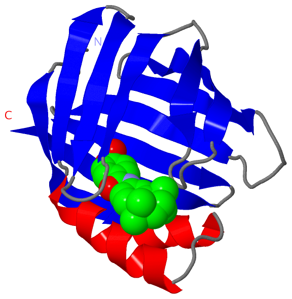
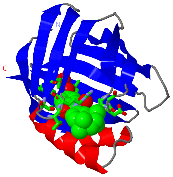
Asymmetric/Biological Unit(hide GO term definitions)
Chain A   (RABP1_BOVIN | P62964)
molecular function
    GO:0008289    lipid binding    Interacting selectively and non-covalently with a lipid.
    GO:0016918    retinal binding    Interacting selectively and non-covalently with retinal, one of the forms of vitamin A. Retinal plays an important role in the visual process in most vertebrates, combining with opsins to form visual pigments in the retina.
    GO:0001972    retinoic acid binding    Interacting selectively and non-covalently with retinoic acid, 3,7-dimethyl-9-(2,6,-trimethyl-1-cyclohexen-1-yl)-2,4,6,8-nonatetraenoic acid.
    GO:0019841    retinol binding    Interacting selectively and non-covalently with retinol, vitamin A1, 2,6,6-trimethyl-1-(9'-hydroxy-3',7'-dimethylnona-1',3',5',7'-tetraenyl)cyclohex-1-ene, one of the three components that makes up vitamin A. Retinol is an intermediate in the vision cycle and it also plays a role in growth and differentiation.
    GO:0005215    transporter activity    Enables the directed movement of substances (such as macromolecules, small molecules, ions) into, out of or within a cell, or between cells.
biological process
    GO:0034653    retinoic acid catabolic process    The chemical reactions and pathways resulting in the breakdown of retinoic acid, one of the three components that makes up vitamin A.
    GO:0006810    transport    The directed movement of substances (such as macromolecules, small molecules, ions) or cellular components (such as complexes and organelles) into, out of or within a cell, or between cells, or within a multicellular organism by means of some agent such as a transporter, pore or motor protein.
cellular component
    GO:0005737    cytoplasm    All of the contents of a cell excluding the plasma membrane and nucleus, but including other subcellular structures.

 Visualization

(-) Interactive Views

Asymmetric/Biological Unit
  Complete Structure
    Jena3D(integrated viewing of ligand, site, SAP, PROSITE, SCOP information)
    WebMol | AstexViewer[tm]@PDBe
(Java Applets, require no local installation except for Java; loading may be slow)
    STRAP
(Java WebStart application, automatic local installation, requires Java; full application with system access!)
    RasMol
(require local installation)
    Molscript (VRML)
(requires installation of a VRML viewer; select preferred view via VRML and generate a mono or stereo PDF format file)
 
  Ligands, Modified Residues, Ions
    A80  [ RasMol | Jena3D ]  +environment [ RasMol | Jena3D ]
 
  Sites
    AC1  [ RasMol ]  +environment [ RasMol ]
 
  Cis Peptide Bonds
(no "Cis Peptide Bonds" information available for 2cbr)
 

(-) Still Images

Jmol
  protein: cartoon or spacefill or dots and stick; nucleic acid: cartoon and stick; ligands: spacefill; active site: stick
Molscript
  protein, nucleic acid: cartoon; ligands: spacefill; active site: ball and stick

 Databases and Analysis Tools

(-) Databases

Access by PDB/NDB ID
  2cbr
    Family and Domain InformationProDom | SYSTERS
    General Structural InformationGlycoscienceDB | MMDB | NDB | OCA | PDB | PDBe | PDBj | PDBsum | PDBWiki | PQS | PROTEOPEDIA
    Orientation in MembranesOPM
    Protein SurfaceSURFACE
    Secondary StructureDSSP (structure derived) | HSSP (homology derived)
    Structural GenomicsGeneCensus
    Structural NeighboursCE | VAST
    Structure ClassificationCATH | Dali | SCOP
    Validation and Original DataBMRB Data View | BMRB Restraints Grid | EDS | PROCHECK | RECOORD | WHAT_CHECK
 
Access by UniProt ID/Accession number
  RABP1_BOVIN | P62964
    Comparative Protein Structure ModelsModBase
    Genomic InformationEnsembl
    Protein-protein InteractionDIP
    Sequence, Family and Domain InformationInterPro | Pfam | SMART | UniProtKB/SwissProt
 
Access by Enzyme Classificator   (EC Number)
  (no 'Enzyme Classificator' available)
    General Enzyme InformationBRENDA | EC-PDB | Enzyme | IntEnz
    PathwayKEGG | MetaCyc
 
Access by Disease Identifier   (MIM ID)
  (no 'MIM ID' available)
    Disease InformationOMIM
 
Access by GenAge ID
  (no 'GenAge ID' available)
    Age Related InformationGenAge

(-) Analysis Tools

Access by PDB/NDB ID
    Domain InformationXDom
    Interatomic Contacts of Structural UnitsCSU
    Ligand-protein ContactsLPC
    Protein CavitiescastP
    Sequence and Secondary StructurePDBCartoon
    Structure AlignmentSTRAP(Java WebStart application, automatic local installation, requires Java; full application with system access!)
    Structure and Sequence BrowserSTING
 
Access by UniProt ID/Accession number
  RABP1_BOVIN | P62964
    Protein Disorder PredictionDisEMBL | FoldIndex | GLOBPLOT (for more information see DisProt)

 Related Entries

(-) Entries Sharing at Least One Protein Chain (UniProt ID)

(no "Entries Sharing at Least One Protein Chain" available for 2CBR)

(-) Related Entries Specified in the PDB File

(no "Related Entries Specified in the PDB File" available for 2CBR)