|
|
|
|
 Description
|
|
Compounds
|
||||||||||||||||||||||||||||||||||||||||||||||||||||||||
Chains, Units
Summary Information (see also Sequences/Alignments below) |
Ligands, Modified Residues, Ions (0, 0)| (no "Ligand,Modified Residues,Ions" information available for 2BT8) |
Sites (0, 0)| (no "Site" information available for 2BT8) |
SS Bonds (0, 0)| (no "SS Bond" information available for 2BT8) |
Cis Peptide Bonds (1, 1)
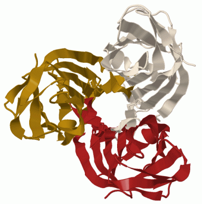
Asymmetric Unit
|
||||||||
SAPs(SNPs)/Variants (0, 0)| (no "SAP(SNP)/Variant" information available for 2BT8) |
PROSITE Motifs (0, 0)| (no "PROSITE Motif" information available for 2BT8) |
Exons (0, 0)| (no "Exon" information available for 2BT8) |
Sequences/Alignments
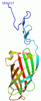
Asymmetric UnitChain A from PDB Type:PROTEIN Length:167 aligned with O12287_9REOV | O12287 from UniProtKB/TrEMBL Length:326 Alignment length:170 166 176 186 196 206 216 226 236 246 256 266 276 286 296 306 316 326 O12287_9REOV 157 SHGLSFSPPLSVADGVVSLDMDPYFCSQRVSLTSYSAEAQLMQFRWMARGTNGSSDTIDMTVNAHCHGRRTDYMMSSTGNLTVTSNVVLLTFDLSDITHIPSDLARLVPSAGFQAASFPVDVSFTRDSATHAYQAYGVYSSSRVFTITFPTGGDGTANIRSLTVRTGIDT 326 SCOP domains -------------------------------------------------------------------------------------------------------------------------------------------------------------------------- SCOP domains CATH domains -------------------------------------------------------------------------------------------------------------------------------------------------------------------------- CATH domains Pfam domains -------------------------------------------------------------------------------------------------------------------------------------------------------------------------- Pfam domains SAPs(SNPs) -------------------------------------------------------------------------------------------------------------------------------------------------------------------------- SAPs(SNPs) PROSITE -------------------------------------------------------------------------------------------------------------------------------------------------------------------------- PROSITE Transcript -------------------------------------------------------------------------------------------------------------------------------------------------------------------------- Transcript 2bt8 A 157 SHGLSFSPPLSVADGVVSLDMDPYFCSQRVSLTSYSAEAQLMQFRWMARGTNGSSDTIDMTVNAHCHGRRTDYMMSSTGNLTVTSNVVLLTFDLSDIT---SDLARLVPSAGFQAASFPVDVSFTRDSATHAYQAYGVYSSSRVFTITFPTGGDGTANIRSLTVRTGIDT 326 166 176 186 196 206 216 226 236 246 | - | 266 276 286 296 306 316 326 254 258 Chain A from PDB Type:PROTEIN Length:167 aligned with SIGC_ARVS1 | Q992I2 from UniProtKB/Swiss-Prot Length:326 Alignment length:170 166 176 186 196 206 216 226 236 246 256 266 276 286 296 306 316 326 SIGC_ARVS1 157 SHGLSFSPPLSVADGVVSLDMDPYFCSQRVSLTSYSAEAQLMQFRWMARGTNGSSDTIDMTVNAHCHGRRTDYMMSSTGNLTVTSNVVLLTFDLSDITHIPSDLARLVPSAGFQAASFPVDVSFTRDSATHAYQAYGVYSSSRVFTITFPTGGDGTANIRSLTVRTGIDT 326 SCOP domains -------------------------------------------------------------------------------------------------------------------------------------------------------------------------- SCOP domains CATH domains -------------------------------------------------------------------------------------------------------------------------------------------------------------------------- CATH domains Pfam domains -------------------------------------------------------------------------------------------------------------------------------------------------------------------------- Pfam domains SAPs(SNPs) -------------------------------------------------------------------------------------------------------------------------------------------------------------------------- SAPs(SNPs) PROSITE -------------------------------------------------------------------------------------------------------------------------------------------------------------------------- PROSITE Transcript -------------------------------------------------------------------------------------------------------------------------------------------------------------------------- Transcript 2bt8 A 157 SHGLSFSPPLSVADGVVSLDMDPYFCSQRVSLTSYSAEAQLMQFRWMARGTNGSSDTIDMTVNAHCHGRRTDYMMSSTGNLTVTSNVVLLTFDLSDIT---SDLARLVPSAGFQAASFPVDVSFTRDSATHAYQAYGVYSSSRVFTITFPTGGDGTANIRSLTVRTGIDT 326 166 176 186 196 206 216 226 236 246 | - | 266 276 286 296 306 316 326 254 258
|
||||||||||||||||||||
SCOP Domains (0, 0)| (no "SCOP Domain" information available for 2BT8) |
CATH Domains (0, 0)| (no "CATH Domain" information available for 2BT8) |
Pfam Domains (0, 0)| (no "Pfam Domain" information available for 2BT8) |
Gene Ontology (5, 5)|
Asymmetric Unit(hide GO term definitions) Chain A (SIGC_ARVS1 | Q992I2)
|
||||||||||||||||||||||||||||||||||||||||||
Interactive Views
|
|||||||||||||||||||||||||||||||||||||||||||||||||||||||||||||||||||||||||||||||||||||||||||||||||||||||||||||||||||||||||||||||||||||||
Still Images
|
||||||||||||||||
Databases
|
||||||||||||||||||||||||||||||||||||||||||||||||||||||||||||||||||||||||||||||||||||||||||||||||||||||||||||||||||||||||||||||||||||||||||||||||||||||||||||||||||||||||||||||||||||||||||
Analysis Tools
|
||||||||||||||||||||||||||||||||||||||||||||||||||||||||||||||||||||||||
Entries Sharing at Least One Protein Chain (UniProt ID)
Related Entries Specified in the PDB File
|
|