|
|
|
|
 Description
|
|
Compounds
|
||||||||||||||||||||||||||||||||||||||||
Chains, Units
Summary Information (see also Sequences/Alignments below) |
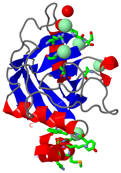
Ligands, Modified Residues, Ions (1, 5)
Asymmetric/Biological Unit (1, 5)
|
Sites (5, 5)
Asymmetric Unit (5, 5)
|
SS Bonds (0, 0)| (no "SS Bond" information available for 2B71) |
Cis Peptide Bonds (0, 0)| (no "Cis Peptide Bond" information available for 2B71) |
SAPs(SNPs)/Variants (0, 0)| (no "SAP(SNP)/Variant" information available for 2B71) |
PROSITE Motifs (0, 0)| (no "PROSITE Motif" information available for 2B71) |
Exons (0, 0)| (no "Exon" information available for 2B71) |
Sequences/Alignments
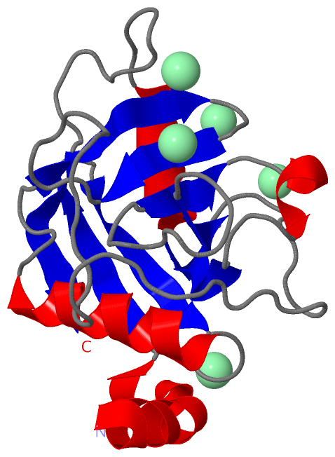
Asymmetric/Biological UnitChain A from PDB Type:PROTEIN Length:169 aligned with Q7RRM6_PLAYO | Q7RRM6 from UniProtKB/TrEMBL Length:202 Alignment length:169 38 48 58 68 78 88 98 108 118 128 138 148 158 168 178 188 Q7RRM6_PLAYO 29 LEEKIAYYKMKGHTERGYITIYTNLGDFEVELYWYHSPKTCLNFYTLCEMGFYDNTIFHRVIPNFVIQGGDPTGTGKGGKSIYGEYFEDEINKELKHTGAGILSMSNNGPNTNSSQFFITLAPLPHLDGKHTIFARVSKNMTCIENIASVQTTATNKPIFDLKILRTST 197 SCOP domains d2b71a1 A:23-191 Cyclophilin-like protein PY00693 SCOP domains CATH domains 2b71A00 A:23-191 Cyclophilin CATH domains Pfam domains ------------------------------------------------------------------------------------------------------------------------------------------------------------------------- Pfam domains SAPs(SNPs) ------------------------------------------------------------------------------------------------------------------------------------------------------------------------- SAPs(SNPs) PROSITE ------------------------------------------------------------------------------------------------------------------------------------------------------------------------- PROSITE Transcript ------------------------------------------------------------------------------------------------------------------------------------------------------------------------- Transcript 2b71 A 23 LEEKIAYYKMKGHTERGYITIYTNLGDFEVELYWYHSPKTCLNFYTLCEMGFYDNTIFHRVIPNFVIQGGDPTGTGKGGKSIYGEYFEDEINKELKHTGAGILSMSNNGPNTNSSQFFITLAPLPHLDGKHTIFARVSKNMTCIENIASVQTTATNKPIFDLKILRTST 191 32 42 52 62 72 82 92 102 112 122 132 142 152 162 172 182
|
||||||||||||||||||||
SCOP Domains (1, 1)
Asymmetric/Biological Unit
 |
CATH Domains (1, 1)
Asymmetric/Biological Unit
|
Pfam Domains (0, 0)| (no "Pfam Domain" information available for 2B71) |
Gene Ontology (4, 4)|
Asymmetric/Biological Unit(hide GO term definitions) Chain A (Q7RRM6_PLAYO | Q7RRM6)
|
||||||||||||||||||||||||||||||||||||
Interactive Views
|
||||||||||||||||||||||||||||||||||||||||||||||||||||||||||||||||||||||||||||||||||||||||||||||||||||||||||||||||||||||||||||||||||||||||||||||||||
Still Images
|
||||||||||||||||
Databases
|
||||||||||||||||||||||||||||||||||||||||||||||||||||||||||||||||||||||||||||||||||||||||||||||||||||||||||||||||||||||||||||||||||||||||||||||||||||||||||||||||
Analysis Tools
|
|||||||||||||||||||||||||||||||||||||||||||||||||||||||||||||
Entries Sharing at Least One Protein Chain (UniProt ID)
Related Entries Specified in the PDB File
|
|