Show PDB file:   
         Plain Text   HTML   (compressed file size)
QuickSearch:   
by PDB,NDB,UniProt,PROSITE Code or Search Term(s)  
(-)Asym./Biol. Unit - manually
(-)Asym./Biol. Unit
(-)Asym./Biol. Unit - sites
collapse expand < >
Image Asym./Biol. Unit - manually
Asym./Biol. Unit - manually  (Jmol Viewer)
Image Asym./Biol. Unit
Asym./Biol. Unit  (Jmol Viewer)
Image Asym./Biol. Unit - sites
Asym./Biol. Unit - sites  (Jmol Viewer)

(-) Description

Title :  THE 1.8 A CRYSTAL STRUCTURE OF A PROTEINASE K LIKE ENZYME FROM A PSYCHROTROPH SERRATIA SPECIES
 
Authors :  R. Helland, A. N. Larsen, A. O. Smalas, N. P. Willassen
Date :  03 Oct 05  (Deposition) - 07 Mar 06  (Release) - 13 Jul 11  (Revision)
Method :  X-RAY DIFFRACTION
Resolution :  1.80
Chains :  Asym./Biol. Unit :  A,B
Keywords :  S Binding, Substrate Specificity, Proteinase K, Subtilase, Psychrotrophic, Psychrophilic, Hydrolase (Keyword Search: [Gene Ontology, PubMed, Web (Google))
 
Reference :  R. Helland, A. N. Larsen, A. O. Smalas, N. P. Willassen
The 1. 8 A Crystal Structure Of A Proteinase K-Like Enzyme From A Psychrotroph Serratia Species
Febs J. V. 273 61 2006
PubMed-ID: 16367748  |  Reference-DOI: 10.1111/J.1742-4658.2005.05040.X

(-) Compounds

Molecule 1 - PROTEINASE K
    ChainsA
    EC Number3.4.21.-
    EngineeredYES
    Expression SystemESCHERICHIA COLI
    Expression System PlasmidPBAD-GIII
    Expression System StrainTOP10
    Expression System Taxid562
    Expression System Vector TypePLASMID
    FragmentRESIDUES 1-277
    Organism ScientificSERRATIA SP.
    Organism Taxid348903
    StrainGF96
 
Molecule 2 - TRIPEPTIDE
    ChainsB
    EngineeredYES
    Expression SystemESCHERICHIA COLI
    Expression System Taxid562
    Organism ScientificSERRATIA SP.
    Organism Taxid348903
    Other DetailsTHE PEPTIDE IS ASSUMED TO BE A PROTEOLYSIS PRODUCT EITHER FROM THE EXPRESSION OR PURIFICATION PROCESS.
    StrainGF96

 Structural Features

(-) Chains, Units

  12
Asymmetric/Biological Unit AB

Summary Information (see also Sequences/Alignments below)
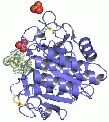
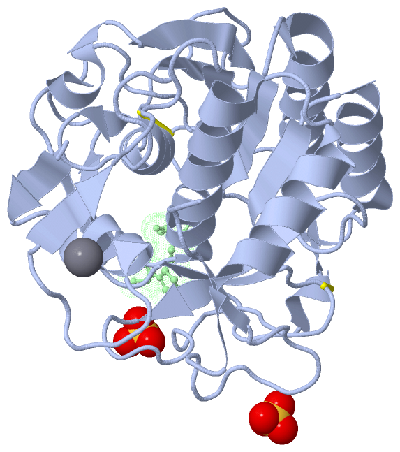
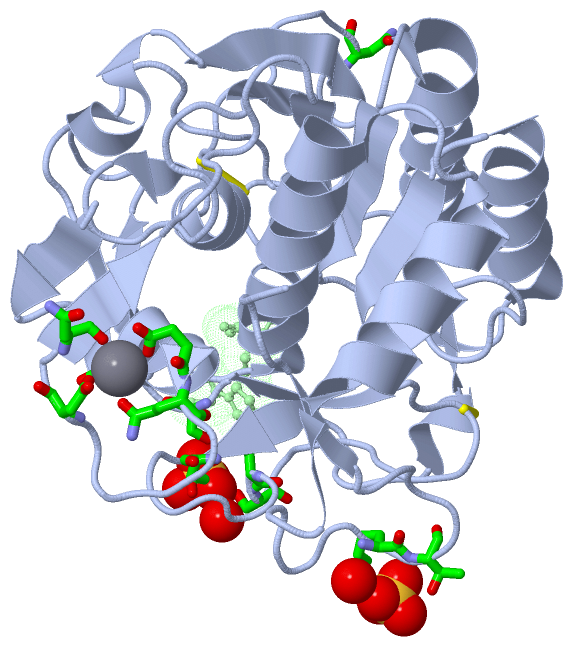
(-) Ligands, Modified Residues, Ions  (2, 3)

Asymmetric/Biological Unit (2, 3)
No.NameCountTypeFull Name
1CA1Ligand/IonCALCIUM ION
2SO42Ligand/IonSULFATE ION

(-) Sites  (3, 3)

Asymmetric Unit (3, 3)
No.NameEvidenceResiduesDescription
1AC1SOFTWAREASP A:11 , ASP A:14 , GLN A:15 , ASP A:21 , ASN A:23 , HOH A:432BINDING SITE FOR RESIDUE CA A 300
2AC2SOFTWARETHR A:184 , SER A:190 , SER A:191 , SER A:206 , HOH A:548BINDING SITE FOR RESIDUE SO4 A 301
3AC3SOFTWAREASN A:60C , LYS A:265 , THR A:266 , HOH A:423BINDING SITE FOR RESIDUE SO4 A 302

(-) SS Bonds  (2, 2)

Asymmetric/Biological Unit
No.Residues
1A:66 -A:98
2A:167 -A:198

(-) Cis Peptide Bonds  (2, 2)

Asymmetric/Biological Unit
No.Residues
1Leu A:18 -Pro A:19
2Ser A:170 -Pro A:171

 Sequence-Structure Mapping

(-) SAPs(SNPs)/Variants  (0, 0)

(no "SAP(SNP)/Variant" information available for 2B6N)

(-) PROSITE Motifs  (0, 0)

(no "PROSITE Motif" information available for 2B6N)

(-) Exons   (0, 0)

(no "Exon" information available for 2B6N)

(-) Sequences/Alignments

Asymmetric/Biological Unit
   Reformat: Number of residues per line =  ('0' or empty: single-line sequence representation)
  Number of residues per labelling interval =   
  UniProt sequence: complete  aligned part    
   Show mapping: SCOP domains CATH domains Pfam domains Secondary structure (by author)
SAPs(SNPs) PROSITE motifs Exons
(details for a mapped element are shown in a popup box when the mouse pointer rests over it)
Chain A from PDB  Type:PROTEIN  Length:278
 aligned with Q3HUQ2_9GAMM | Q3HUQ2 from UniProtKB/TrEMBL  Length:629

    Alignment length:278
                                   136       146       156       166       176       186       196       206       216       226       236       246       256       266       276       286       296       306       316       326       336       346       356       366       376       386       396        
        Q3HUQ2_9GAMM    127 ADQPSPTWGIDRIDQRNLPLDNNYHTDYDGSGVTAFVIDTGVLNTHNEFGGRASSGYDFIDNDYDATDCNGHGTHVAGTIGGSTYGVAKNVNVVGVRVLNCSGSGSNSGVIAGINWVKNNASGPAVANMSLGGGASQATDDAVNAAVAAGITFVVAAGNDNSNACNYSPARAADAITVGSTTSNDSRSSFSNYGTCLDIYAPGSSITSSWYTSNSATNTISGTSMASPHVAGVAALYLDENPNLSPAQVTNLLKTRATADKVTDAKTGSPNKLLFSLA  404
               SCOP domains d2b6na_ A: automated matches                                                                                                                                                                                                                                                           SCOP domains
               CATH domains 2b6nA00 A:1-277  [code=3.40.50.200, no name defined]                                                                                                                                                                                                                                   CATH domains
               Pfam domains -------------------------------------------------------------------------------------------------------------------------------------------------------------------------------------------------------------------------------------------------------------------------------------- Pfam domains
         Sec.struct. author .eee..hhhhhhhh........eee........eeeeee......hhhhh..eeeeee............hhhhhhhhhhhh.........eeeeee........hhhhhhhhhhhhhhhh...eeeee...ee.hhhhhhhhhhhhhh..eeeee......hhh.ee.......eeeeee.................eeee...eeee.......eeee.hhhhhhhhhhhhhhhhhhhh...hhhhhhhhhhhhhee............ee..... Sec.struct. author
                 SAPs(SNPs) -------------------------------------------------------------------------------------------------------------------------------------------------------------------------------------------------------------------------------------------------------------------------------------- SAPs(SNPs)
                    PROSITE -------------------------------------------------------------------------------------------------------------------------------------------------------------------------------------------------------------------------------------------------------------------------------------- PROSITE
                 Transcript -------------------------------------------------------------------------------------------------------------------------------------------------------------------------------------------------------------------------------------------------------------------------------------- Transcript
                2b6n A    1 ADQPSPTWGIDRIDQRNLPLDNNYHTDYDGSGVTAFVIDTGVLNTHNEFGGRASSGYDFIDNDYDATDCNGHGTHVAGTIGGSTYGVAKNVNVVGVRVLNCSGSGSNSGVIAGINWVKNNASGPAVANMSLGGGASQATDDAVNAAVAAGITFVVAAGNDNSNACNYSPARAADAITVGSTTSNDSRSSFSNYGTCLDIYAPGSSITSSWYTSNSATNTISGTSMASPHVAGVAALYLDENPNLSPAQVTNLLKTRATADKVTDAKTGSPNKLLFSLA  277
                                    10        20        30        40        50        60|||     67        77        87        97       107       117 ||    132       142       152       162       172       182       192       202       212 ||    220       230       240 |     249       259       269        
                                                                                      60B||                                                        119|                                                                                     213B|                         241B                                    
                                                                                       60C|                                                         125                                                                                      213C                                                                 
                                                                                        60D                                                                                                                                                                                                                       

Chain B from PDB  Type:PROTEIN  Length:3
                                    
               SCOP domains --- SCOP domains
               CATH domains --- CATH domains
               Pfam domains --- Pfam domains
         Sec.struct. author ... Sec.struct. author
                 SAPs(SNPs) --- SAPs(SNPs)
                    PROSITE --- PROSITE
                 Transcript --- Transcript
                2b6n B    1 APT    3

   Legend:   → Mismatch (orange background)
  - → Gap (green background, '-', border residues have a numbering label)
    → Modified Residue (blue background, lower-case, 'x' indicates undefined single-letter code, labelled with number + name)
  x → Chemical Group (purple background, 'x', labelled with number + name, e.g. ACE or NH2)
  extra numbering lines below/above indicate numbering irregularities and modified residue names etc., number ends below/above '|'

 Classification and Annotation

(-) SCOP Domains  (1, 1)

Asymmetric/Biological Unit

(-) CATH Domains  (1, 1)

Asymmetric/Biological Unit
(-)
Class: Alpha Beta (26913)

(-) Pfam Domains  (0, 0)

(no "Pfam Domain" information available for 2B6N)

(-) Gene Ontology  (6, 6)

Asymmetric/Biological Unit(hide GO term definitions)
Chain A   (Q3HUQ2_9GAMM | Q3HUQ2)
molecular function
    GO:0016787    hydrolase activity    Catalysis of the hydrolysis of various bonds, e.g. C-O, C-N, C-C, phosphoric anhydride bonds, etc. Hydrolase is the systematic name for any enzyme of EC class 3.
    GO:0046872    metal ion binding    Interacting selectively and non-covalently with any metal ion.
    GO:0008233    peptidase activity    Catalysis of the hydrolysis of a peptide bond. A peptide bond is a covalent bond formed when the carbon atom from the carboxyl group of one amino acid shares electrons with the nitrogen atom from the amino group of a second amino acid.
    GO:0004252    serine-type endopeptidase activity    Catalysis of the hydrolysis of internal, alpha-peptide bonds in a polypeptide chain by a catalytic mechanism that involves a catalytic triad consisting of a serine nucleophile that is activated by a proton relay involving an acidic residue (e.g. aspartate or glutamate) and a basic residue (usually histidine).
    GO:0008236    serine-type peptidase activity    Catalysis of the hydrolysis of peptide bonds in a polypeptide chain by a catalytic mechanism that involves a catalytic triad consisting of a serine nucleophile that is activated by a proton relay involving an acidic residue (e.g. aspartate or glutamate) and a basic residue (usually histidine).
biological process
    GO:0006508    proteolysis    The hydrolysis of proteins into smaller polypeptides and/or amino acids by cleavage of their peptide bonds.

 Visualization

(-) Interactive Views

Asymmetric/Biological Unit
  Complete Structure
    Jena3D(integrated viewing of ligand, site, SAP, PROSITE, SCOP information)
    WebMol | AstexViewer[tm]@PDBe
(Java Applets, require no local installation except for Java; loading may be slow)
    STRAP
(Java WebStart application, automatic local installation, requires Java; full application with system access!)
    RasMol
(require local installation)
    Molscript (VRML)
(requires installation of a VRML viewer; select preferred view via VRML and generate a mono or stereo PDF format file)
 
  Ligands, Modified Residues, Ions
    CA  [ RasMol | Jena3D ]  +environment [ RasMol | Jena3D ]
    SO4  [ RasMol | Jena3D ]  +environment [ RasMol | Jena3D ]
 
  Sites
    AC1  [ RasMol ]  +environment [ RasMol ]
    AC2  [ RasMol ]  +environment [ RasMol ]
    AC3  [ RasMol ]  +environment [ RasMol ]
 
  Cis Peptide Bonds
    Leu A:18 - Pro A:19   [ RasMol ]  
    Ser A:170 - Pro A:171   [ RasMol ]  
 

(-) Still Images

Jmol
  protein: cartoon or spacefill or dots and stick; nucleic acid: cartoon and stick; ligands: spacefill; active site: stick
Molscript
  protein, nucleic acid: cartoon; ligands: spacefill; active site: ball and stick

 Databases and Analysis Tools

(-) Databases

Access by PDB/NDB ID
  2b6n
    Family and Domain InformationProDom | SYSTERS
    General Structural InformationGlycoscienceDB | MMDB | NDB | OCA | PDB | PDBe | PDBj | PDBsum | PDBWiki | PQS | PROTEOPEDIA
    Orientation in MembranesOPM
    Protein SurfaceSURFACE
    Secondary StructureDSSP (structure derived) | HSSP (homology derived)
    Structural GenomicsGeneCensus
    Structural NeighboursCE | VAST
    Structure ClassificationCATH | Dali | SCOP
    Validation and Original DataBMRB Data View | BMRB Restraints Grid | EDS | PROCHECK | RECOORD | WHAT_CHECK
 
Access by UniProt ID/Accession number
  Q3HUQ2_9GAMM | Q3HUQ2
    Comparative Protein Structure ModelsModBase
    Genomic InformationEnsembl
    Protein-protein InteractionDIP
    Sequence, Family and Domain InformationInterPro | Pfam | SMART | UniProtKB/TrEMBL
 
Access by Enzyme Classificator   (EC Number)
  3.4.21.-
    General Enzyme InformationBRENDA | EC-PDB | Enzyme | IntEnz
    PathwayKEGG | MetaCyc
 
Access by Disease Identifier   (MIM ID)
  (no 'MIM ID' available)
    Disease InformationOMIM
 
Access by GenAge ID
  (no 'GenAge ID' available)
    Age Related InformationGenAge

(-) Analysis Tools

Access by PDB/NDB ID
    Domain InformationXDom
    Interatomic Contacts of Structural UnitsCSU
    Ligand-protein ContactsLPC
    Protein CavitiescastP
    Sequence and Secondary StructurePDBCartoon
    Structure AlignmentSTRAP(Java WebStart application, automatic local installation, requires Java; full application with system access!)
    Structure and Sequence BrowserSTING
 
Access by UniProt ID/Accession number
  Q3HUQ2_9GAMM | Q3HUQ2
    Protein Disorder PredictionDisEMBL | FoldIndex | GLOBPLOT (for more information see DisProt)

 Related Entries

(-) Entries Sharing at Least One Protein Chain (UniProt ID)

(no "Entries Sharing at Least One Protein Chain" available for 2B6N)

(-) Related Entries Specified in the PDB File

(no "Related Entries Specified in the PDB File" available for 2B6N)