|
|
|
|
 Description
|
|
Compounds
|
||||||||||||||||||||||||||||||||||||
Chains, Units
Summary Information (see also Sequences/Alignments below) |
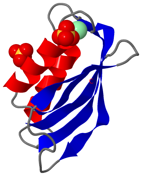
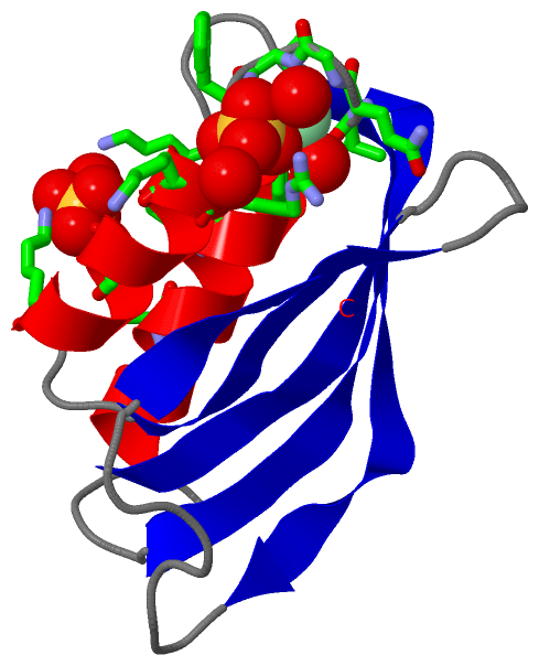
Ligands, Modified Residues, Ions (2, 3)| Asymmetric/Biological Unit (2, 3) |
Sites (3, 3)
Asymmetric Unit (3, 3)
|
SS Bonds (0, 0)| (no "SS Bond" information available for 2ACY) |
Cis Peptide Bonds (0, 0)| (no "Cis Peptide Bond" information available for 2ACY) |
SAPs(SNPs)/Variants (0, 0)| (no "SAP(SNP)/Variant" information available for 2ACY) |
PROSITE Motifs (3, 3)
Asymmetric/Biological Unit (3, 3)
|
||||||||||||||||||||||||||||||||||||||||
Exons (2, 2)
Asymmetric/Biological Unit (2, 2)
|
||||||||||||||||||||||||||||||||||||||||||||||||||||||||||||
Sequences/Alignments
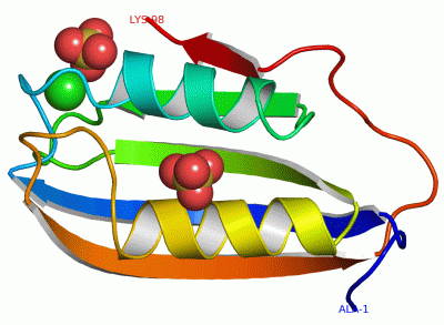
Asymmetric/Biological UnitChain A from PDB Type:PROTEIN Length:98 aligned with ACYP1_BOVIN | P41500 from UniProtKB/Swiss-Prot Length:101 Alignment length:98 13 23 33 43 53 63 73 83 93 ACYP1_BOVIN 4 AEGDTLISVDYEIFGKVQGVFFRKYTQAEGKKLGLVGWVQNTDQGTVQGQLQGPASKVRHMQEWLETKGSPKSHIDRASFHNEKVIVKLDYTDFQIVK 101 SCOP domains d2acya_ A: Acylphosphatase SCOP domains CATH domains 2acyA00 A:1-98 [code=3.30.70.100, no name defined] CATH domains Pfam domains -------------------------------------------------------------------------------------------------- Pfam domains SAPs(SNPs) -------------------------------------------------------------------------------------------------- SAPs(SNPs) PROSITE (1) -------ACYLPHOSPHATASE_3 PDB: A:8-98 UniProt: 11-101 PROSITE (1) PROSITE (2) ------------ACYLPHOSPHA-------------ACYLPHOSPHATASE_2--------------------------------------------- PROSITE (2) Transcript 1 Exon 1.2 PDB: A:1-27 Exon 1.3 PDB: A:28-98 UniProt: 31-101 Transcript 1 2acy A 1 AEGDTLISVDYEIFGKVQGVFFRKYTQAEGKKLGLVGWVQNTDQGTVQGQLQGPASKVRHMQEWLETKGSPKSHIDRASFHNEKVIVKLDYTDFQIVK 98 10 20 30 40 50 60 70 80 90
|
||||||||||||||||||||
SCOP Domains (1, 1)
Asymmetric/Biological Unit
 |
CATH Domains (1, 1)
Asymmetric/Biological Unit
|
Pfam Domains (0, 0)| (no "Pfam Domain" information available for 2ACY) |
Gene Ontology (2, 2)|
Asymmetric/Biological Unit(hide GO term definitions) Chain A (ACYP1_BOVIN | P41500)
|
||||||||||||||||||
Interactive Views
|
|||||||||||||||||||||||||||||||||||||||||||||||||||||||||||||||||||||||||||||||||||||||||||||||||||||||||||||||||||||||||||||||||||||||||||
Still Images
|
||||||||||||||||
Databases
|
||||||||||||||||||||||||||||||||||||||||||||||||||||||||||||||||||||||||||||||||||||||||||||||||||||||||||||||||||||||||||||||||||||||||||||||||||||||||||||||||
Analysis Tools
|
|||||||||||||||||||||||||||||||||||||||||||||||||||||||||||||
Entries Sharing at Least One Protein Chain (UniProt ID)
Related Entries Specified in the PDB File
|
|