|
|
|
|
 Description
|
|
Compounds
|
||||||||||||||||||||||||||||||||||||||||||||
Chains, Units
Summary Information (see also Sequences/Alignments below) |
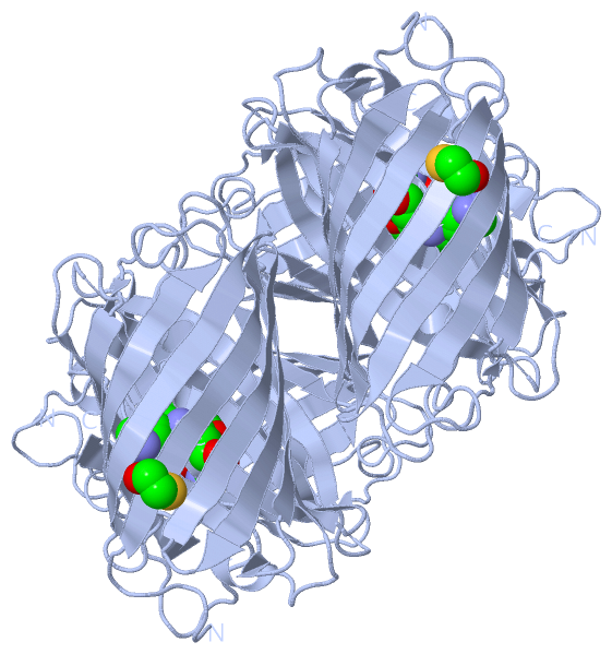
Ligands, Modified Residues, Ions (2, 2)| Asymmetric Unit (2, 2) Biological Unit 1 (2, 8) |
Sites (1, 1)
Asymmetric Unit (1, 1)
|
SS Bonds (0, 0)| (no "SS Bond" information available for 2A47) |
Cis Peptide Bonds (1, 1)
Asymmetric Unit
|
||||||||
SAPs(SNPs)/Variants (0, 0)| (no "SAP(SNP)/Variant" information available for 2A47) |
PROSITE Motifs (0, 0)| (no "PROSITE Motif" information available for 2A47) |
Exons (0, 0)| (no "Exon" information available for 2A47) |
Sequences/Alignments
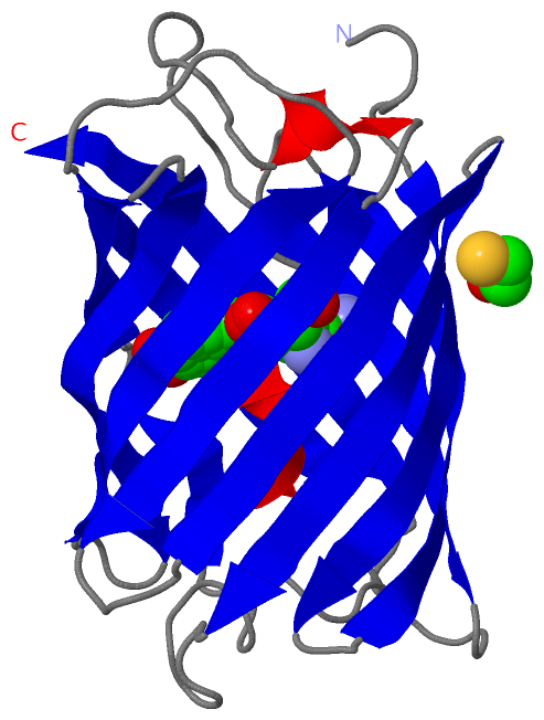
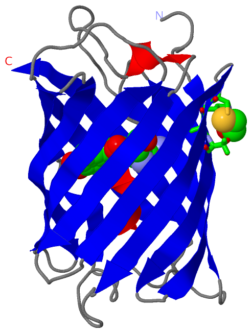
Asymmetric UnitChain A from PDB Type:PROTEIN Length:218 aligned with GFPL_ANEMA | Q9U6Y6 from UniProtKB/Swiss-Prot Length:229 Alignment length:220 14 24 34 44 54 64 74 84 94 104 114 124 134 144 154 164 174 184 194 204 214 224 GFPL_ANEMA 5 NKFIGDDMKMTYHMDGCVNGHYFTVKGEGNGKPYEGTQTSTFKVTMANGGPLAFSFDILSTVFKYGNRCFTAYPTSMPDYFKQAFPDGMSYERTFTYEDGGVATASWEISLKGNCFEHKSTFHGVNFPADGPVMAKKTTGWDPSFEKMTVCDGILKGDVTAFLMLQGGGNYRCQFHTSYKTKKPVTMPPNHVVEHRIARTDLDKGGNSVQLTEHAVAHIT 224 SCOP domains d2a47a_ A: automated matches SCOP domains CATH domains 2a47A00 A:5-224 Green Fluorescent Protein CATH domains Pfam domains ---------------------------------------------------------------------------------------------------------------------------------------------------------------------------------------------------------------------------- Pfam domains SAPs(SNPs) ---------------------------------------------------------------------------------------------------------------------------------------------------------------------------------------------------------------------------- SAPs(SNPs) PROSITE ---------------------------------------------------------------------------------------------------------------------------------------------------------------------------------------------------------------------------- PROSITE Transcript ---------------------------------------------------------------------------------------------------------------------------------------------------------------------------------------------------------------------------- Transcript 2a47 A 5 NKFIGDDMKMTYHMDGCVNGHYFTVKGEGNGKPYEGTQTSTFKVTMANGGPLAFSFDILSTVFk--NRCFTAYPTSMPDYFKQAFPDGMSYERTFTYEDGGVATASWEISLKGNCFEHKSTFHGVNFPADGPVMAKKTTGWDPSFEKMTVCDGILKGDVTAFLMLQGGGNYRCQFHTSYKTKKPVTMPPNHVVETRIARTDLDKGGNSVQLTEHAVAHIT 224 14 24 34 44 54 64 | | 74 84 94 104 114 124 134 144 154 164 174 184 194 204 214 224 68-CR7
|
||||||||||||||||||||
SCOP Domains (1, 1)
Asymmetric Unit
 |
CATH Domains (1, 1)
Asymmetric Unit
|
Pfam Domains (0, 0)| (no "Pfam Domain" information available for 2A47) |
Gene Ontology (3, 3)|
Asymmetric Unit(hide GO term definitions) Chain A (GFPL_ANEMA | Q9U6Y6)
|
||||||||||||||||||||||||
Interactive Views
|
||||||||||||||||||||||||||||||||||||||||||||||||||||||||||||||||||||||||||||||||||||||||||||||||||||||||||||||||||||||||||||||||||||||||||||||||
Still Images
|
||||||||||||||||
Databases
|
||||||||||||||||||||||||||||||||||||||||||||||||||||||||||||||||||||||||||||||||||||||||||||||||||||||||||||||||||||||||||||||||||||||||||||||||||||||||||||||||
Analysis Tools
|
|||||||||||||||||||||||||||||||||||||||||||||||||||||||||||||
Entries Sharing at Least One Protein Chain (UniProt ID)
Related Entries Specified in the PDB File
|
|