Show PDB file:   
         Plain Text   HTML   (compressed file size)
QuickSearch:   
by PDB,NDB,UniProt,PROSITE Code or Search Term(s)  
(-)Asym./Biol. Unit - manually
(-)Asym./Biol. Unit
(-)Asym./Biol. Unit - sites
collapse expand < >
Image Asym./Biol. Unit - manually
Asym./Biol. Unit - manually  (Jmol Viewer)
Image Asym./Biol. Unit
Asym./Biol. Unit  (Jmol Viewer)
Image Asym./Biol. Unit - sites
Asym./Biol. Unit - sites  (Jmol Viewer)

(-) Description

Title :  ADENYLATION DOMAIN OF NAD+ DEPENDENT DNA LIGASE FROM M.TUBERCULOSIS
 
Authors :  S. K. Srivastava, R. Ramachandran
Date :  07 Apr 05  (Deposition) - 05 Jul 05  (Release) - 24 Feb 09  (Revision)
Method :  X-RAY DIFFRACTION
Resolution :  3.15
Chains :  Asym./Biol. Unit :  A
Keywords :  Nad+ Dependent Dna Ligase, Amp, M. Tuberculosis (Keyword Search: [Gene Ontology, PubMed, Web (Google))
 
Reference :  S. K. Srivastava, R. P. Tripathi, R. Ramachandran
Nad+-Dependent Dna Ligase (Rv3014C) From Mycobacterium Tuberculosis: Crystal Structure Of The Adenylation Domain And Identification Of Novel Inhibitors
J. Biol. Chem. V. 280 30273 2005
PubMed-ID: 15901723  |  Reference-DOI: 10.1074/JBC.M503780200
(for further references see the PDB file header)

(-) Compounds

Molecule 1 - DNA LIGASE
    ChainsA
    EC Number6.5.1.2
    EngineeredYES
    Expression SystemESCHERICHIA COLI BL21(DE3)
    Expression System PlasmidPQE60
    Expression System StrainBL21 (DE3)
    Expression System Taxid469008
    Expression System Vector TypePLASMID
    FragmentADENYLATION DOMAIN (RESIDUES 1-328)
    GeneLIGA, LIG
    Organism ScientificMYCOBACTERIUM TUBERCULOSIS
    Organism Taxid1773
    SynonymPOLYDEOXYRIBONUCLEOTIDE SYNTHASE [NAD+]

 Structural Features

(-) Chains, Units

  1
Asymmetric/Biological Unit A

Summary Information (see also Sequences/Alignments below)

(-) Ligands, Modified Residues, Ions  (1, 1)

Asymmetric/Biological Unit (1, 1)
No.NameCountTypeFull Name
1AMP1Ligand/IonADENOSINE MONOPHOSPHATE

(-) Sites  (1, 1)

Asymmetric Unit (1, 1)
No.NameEvidenceResiduesDescription
1AC1SOFTWARELEU A:90 , SER A:91 , LEU A:92 , ASN A:94 , GLU A:121 , LEU A:122 , LYS A:123 , ILE A:124 , ALA A:128 , GLU A:184 , HIS A:236 , LYS A:300BINDING SITE FOR RESIDUE AMP A 400

(-) SS Bonds  (0, 0)

(no "SS Bond" information available for 1ZAU)

(-) Cis Peptide Bonds  (0, 0)

(no "Cis Peptide Bond" information available for 1ZAU)

 Sequence-Structure Mapping

(-) SAPs(SNPs)/Variants  (0, 0)

(no "SAP(SNP)/Variant" information available for 1ZAU)

(-) PROSITE Motifs  (1, 1)

Asymmetric/Biological Unit (1, 1)
 PROSITEUniProtKBPDB
No.IDACDescriptionIDLocationCountLocation
1DNA_LIGASE_N1PS01055 NAD-dependent DNA ligase signature 1.DNLJ_MYCTO123-152  1A:123-152
DNLJ_MYCTU123-152  1A:123-152

(-) Exons   (0, 0)

(no "Exon" information available for 1ZAU)

(-) Sequences/Alignments

Asymmetric/Biological Unit
   Reformat: Number of residues per line =  ('0' or empty: single-line sequence representation)
  Number of residues per labelling interval =   
  UniProt sequence: complete  aligned part    
   Show mapping: SCOP domains CATH domains Pfam domains Secondary structure (by author)
SAPs(SNPs) PROSITE motifs Exons
(details for a mapped element are shown in a popup box when the mouse pointer rests over it)
Chain A from PDB  Type:PROTEIN  Length:321
 aligned with DNLJ_MYCTO | P9WNV0 from UniProtKB/Swiss-Prot  Length:691

    Alignment length:321
                                    17        27        37        47        57        67        77        87        97       107       117       127       137       147       157       167       177       187       197       207       217       227       237       247       257       267       277       287       297       307       317       327 
           DNLJ_MYCTO     8 QTAPEVLRQWQALAEEVREHQFRYYVRDAPIISDAEFDELLRRLEALEEQHPELRTPDSPTQLVGGAGFATDFEPVDHLERMLSLDNAFTADELAAWAGRIHAEVGDAAHYLCELKIDGVALSLVYREGRLTRASTRGDGRTGEDVTLNARTIADVPERLTPGDDYPVPEVLEVRGEVFFRLDDFQALNASLVEEGKAPFANPRNSAAGSLRQKDPAVTARRRLRMICHGLGHVEGFRPATLHQAYLALRAWGLPVSEHTTLATDLAGVRERIDYWGEHRHEVDHEIDGVVVKVDEVALQRRLGSTSRAPRWAIAYKYPPE 328
               SCOP domains d1zaua_ A: automated matches                                                                                                                                                                                                                                                                                                      SCOP domains
               CATH domains 1zauA01 A:8-77 Helix hairpin bin                                      ----------------------------------------------------------------------------------------------------------------------------------------------------------------------------------------------------------------------------------------------------------- CATH domains
               Pfam domains ---DNA_ligase_aden-1zauA01 A:11-328                                                                                                                                                                                                                                                                                               Pfam domains
         Sec.struct. author .hhhhhhhhhhhhhhhhhhhhhhhh........hhhhhhhhhhhhhhhhh.........................ee............hhhhhhhhhhhhhh.......eeeeeee..eeeeee.hhhh...........ee..hhhhhhhh...................eeeeee..hhhhhhhhhhh..........hhhhhhhhhhh..hhhhhhhh..................hhhhhhhhhhh............hhhhhhhhhhhhh..........eeeeeeee.hhhhhh..............ee.... Sec.struct. author
                 SAPs(SNPs) --------------------------------------------------------------------------------------------------------------------------------------------------------------------------------------------------------------------------------------------------------------------------------------------------------------------------------- SAPs(SNPs)
                    PROSITE -------------------------------------------------------------------------------------------------------------------DNA_LIGASE_N1  PDB: A:123-152 -------------------------------------------------------------------------------------------------------------------------------------------------------------------------------- PROSITE
                 Transcript --------------------------------------------------------------------------------------------------------------------------------------------------------------------------------------------------------------------------------------------------------------------------------------------------------------------------------- Transcript
                 1zau A   8 QTAPEVLRQWQALAEEVREHQFRYYVRDAPIISDAEFDELLRRLEALEEQHPELRTPDSPTQLVGGAGFATDFEPVDHLERMLSLDNAFTADELAAWAGRIHAEVGDAAHYLCELKIDGVALSLVYREGRLTRASTRGDGRTGEDVTLNARTIADVPERLTPGDDYPVPEVLEVRGEVFFRLDDFQALNASLVEEGKAPFANPRNSAAGSLRQKDPAVTARRRLRMICHGLGHVEGFRPATLHQAYLALRAWGLPVSEHTTLATDLAGVRERIDYWGEHRHEVDHEIDGVVVKVDEVALQRRLGSTSRAPRWAIAYKYPPE 328
                                    17        27        37        47        57        67        77        87        97       107       117       127       137       147       157       167       177       187       197       207       217       227       237       247       257       267       277       287       297       307       317       327 

Chain A from PDB  Type:PROTEIN  Length:321
 aligned with DNLJ_MYCTU | P9WNV1 from UniProtKB/Swiss-Prot  Length:691

    Alignment length:321
                                    17        27        37        47        57        67        77        87        97       107       117       127       137       147       157       167       177       187       197       207       217       227       237       247       257       267       277       287       297       307       317       327 
           DNLJ_MYCTU     8 QTAPEVLRQWQALAEEVREHQFRYYVRDAPIISDAEFDELLRRLEALEEQHPELRTPDSPTQLVGGAGFATDFEPVDHLERMLSLDNAFTADELAAWAGRIHAEVGDAAHYLCELKIDGVALSLVYREGRLTRASTRGDGRTGEDVTLNARTIADVPERLTPGDDYPVPEVLEVRGEVFFRLDDFQALNASLVEEGKAPFANPRNSAAGSLRQKDPAVTARRRLRMICHGLGHVEGFRPATLHQAYLALRAWGLPVSEHTTLATDLAGVRERIDYWGEHRHEVDHEIDGVVVKVDEVALQRRLGSTSRAPRWAIAYKYPPE 328
               SCOP domains d1zaua_ A: automated matches                                                                                                                                                                                                                                                                                                      SCOP domains
               CATH domains 1zauA01 A:8-77 Helix hairpin bin                                      ----------------------------------------------------------------------------------------------------------------------------------------------------------------------------------------------------------------------------------------------------------- CATH domains
               Pfam domains ---DNA_ligase_aden-1zauA01 A:11-328                                                                                                                                                                                                                                                                                               Pfam domains
         Sec.struct. author .hhhhhhhhhhhhhhhhhhhhhhhh........hhhhhhhhhhhhhhhhh.........................ee............hhhhhhhhhhhhhh.......eeeeeee..eeeeee.hhhh...........ee..hhhhhhhh...................eeeeee..hhhhhhhhhhh..........hhhhhhhhhhh..hhhhhhhh..................hhhhhhhhhhh............hhhhhhhhhhhhh..........eeeeeeee.hhhhhh..............ee.... Sec.struct. author
                 SAPs(SNPs) --------------------------------------------------------------------------------------------------------------------------------------------------------------------------------------------------------------------------------------------------------------------------------------------------------------------------------- SAPs(SNPs)
                PROSITE (2) -------------------------------------------------------------------------------------------------------------------DNA_LIGASE_N1  PDB: A:123-152 -------------------------------------------------------------------------------------------------------------------------------------------------------------------------------- PROSITE (2)
                 Transcript --------------------------------------------------------------------------------------------------------------------------------------------------------------------------------------------------------------------------------------------------------------------------------------------------------------------------------- Transcript
                 1zau A   8 QTAPEVLRQWQALAEEVREHQFRYYVRDAPIISDAEFDELLRRLEALEEQHPELRTPDSPTQLVGGAGFATDFEPVDHLERMLSLDNAFTADELAAWAGRIHAEVGDAAHYLCELKIDGVALSLVYREGRLTRASTRGDGRTGEDVTLNARTIADVPERLTPGDDYPVPEVLEVRGEVFFRLDDFQALNASLVEEGKAPFANPRNSAAGSLRQKDPAVTARRRLRMICHGLGHVEGFRPATLHQAYLALRAWGLPVSEHTTLATDLAGVRERIDYWGEHRHEVDHEIDGVVVKVDEVALQRRLGSTSRAPRWAIAYKYPPE 328
                                    17        27        37        47        57        67        77        87        97       107       117       127       137       147       157       167       177       187       197       207       217       227       237       247       257       267       277       287       297       307       317       327 

   Legend:   → Mismatch (orange background)
  - → Gap (green background, '-', border residues have a numbering label)
    → Modified Residue (blue background, lower-case, 'x' indicates undefined single-letter code, labelled with number + name)
  x → Chemical Group (purple background, 'x', labelled with number + name, e.g. ACE or NH2)
  extra numbering lines below/above indicate numbering irregularities and modified residue names etc., number ends below/above '|'

 Classification and Annotation

(-) SCOP Domains  (1, 1)

Asymmetric/Biological Unit

(-) CATH Domains  (1, 1)

Asymmetric/Biological Unit

(-) Pfam Domains  (1, 1)

Asymmetric/Biological Unit

(-) Gene Ontology  (14, 22)

Asymmetric/Biological Unit(hide GO term definitions)
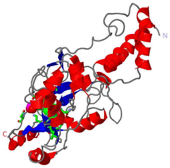
Chain A   (DNLJ_MYCTO | P9WNV0)
molecular function
    GO:0003911    DNA ligase (NAD+) activity    Catalysis of the reaction: NAD+ + deoxyribonucleotide(n) + deoxyribonucleotide(m) = AMP + nicotinamide nucleotide + deoxyribonucleotide(n+m).
    GO:0016874    ligase activity    Catalysis of the joining of two substances, or two groups within a single molecule, with the concomitant hydrolysis of the diphosphate bond in ATP or a similar triphosphate.
    GO:0046872    metal ion binding    Interacting selectively and non-covalently with any metal ion.
biological process
    GO:0006266    DNA ligation    The re-formation of a broken phosphodiester bond in the DNA backbone, carried out by DNA ligase.
    GO:0006259    DNA metabolic process    Any cellular metabolic process involving deoxyribonucleic acid. This is one of the two main types of nucleic acid, consisting of a long, unbranched macromolecule formed from one, or more commonly, two, strands of linked deoxyribonucleotides.
    GO:0006281    DNA repair    The process of restoring DNA after damage. Genomes are subject to damage by chemical and physical agents in the environment (e.g. UV and ionizing radiations, chemical mutagens, fungal and bacterial toxins, etc.) and by free radicals or alkylating agents endogenously generated in metabolism. DNA is also damaged because of errors during its replication. A variety of different DNA repair pathways have been reported that include direct reversal, base excision repair, nucleotide excision repair, photoreactivation, bypass, double-strand break repair pathway, and mismatch repair pathway.
    GO:0006260    DNA replication    The cellular metabolic process in which a cell duplicates one or more molecules of DNA. DNA replication begins when specific sequences, known as origins of replication, are recognized and bound by initiation proteins, and ends when the original DNA molecule has been completely duplicated and the copies topologically separated. The unit of replication usually corresponds to the genome of the cell, an organelle, or a virus. The template for replication can either be an existing DNA molecule or RNA.
    GO:0006974    cellular response to DNA damage stimulus    Any process that results in a change in state or activity of a cell (in terms of movement, secretion, enzyme production, gene expression, etc.) as a result of a stimulus indicating damage to its DNA from environmental insults or errors during metabolism.

Chain A   (DNLJ_MYCTU | P9WNV1)
molecular function
    GO:0003911    DNA ligase (NAD+) activity    Catalysis of the reaction: NAD+ + deoxyribonucleotide(n) + deoxyribonucleotide(m) = AMP + nicotinamide nucleotide + deoxyribonucleotide(n+m).
    GO:0016874    ligase activity    Catalysis of the joining of two substances, or two groups within a single molecule, with the concomitant hydrolysis of the diphosphate bond in ATP or a similar triphosphate.
    GO:0000287    magnesium ion binding    Interacting selectively and non-covalently with magnesium (Mg) ions.
    GO:0046872    metal ion binding    Interacting selectively and non-covalently with any metal ion.
biological process
    GO:0006266    DNA ligation    The re-formation of a broken phosphodiester bond in the DNA backbone, carried out by DNA ligase.
    GO:0006259    DNA metabolic process    Any cellular metabolic process involving deoxyribonucleic acid. This is one of the two main types of nucleic acid, consisting of a long, unbranched macromolecule formed from one, or more commonly, two, strands of linked deoxyribonucleotides.
    GO:0006281    DNA repair    The process of restoring DNA after damage. Genomes are subject to damage by chemical and physical agents in the environment (e.g. UV and ionizing radiations, chemical mutagens, fungal and bacterial toxins, etc.) and by free radicals or alkylating agents endogenously generated in metabolism. DNA is also damaged because of errors during its replication. A variety of different DNA repair pathways have been reported that include direct reversal, base excision repair, nucleotide excision repair, photoreactivation, bypass, double-strand break repair pathway, and mismatch repair pathway.
    GO:0006260    DNA replication    The cellular metabolic process in which a cell duplicates one or more molecules of DNA. DNA replication begins when specific sequences, known as origins of replication, are recognized and bound by initiation proteins, and ends when the original DNA molecule has been completely duplicated and the copies topologically separated. The unit of replication usually corresponds to the genome of the cell, an organelle, or a virus. The template for replication can either be an existing DNA molecule or RNA.
    GO:0006288    base-excision repair, DNA ligation    The ligation by DNA ligase of DNA strands. Ligation occurs after polymerase action to fill the gap left by the action of endonucleases during base-excision repair.
    GO:0006974    cellular response to DNA damage stimulus    Any process that results in a change in state or activity of a cell (in terms of movement, secretion, enzyme production, gene expression, etc.) as a result of a stimulus indicating damage to its DNA from environmental insults or errors during metabolism.
    GO:0040007    growth    The increase in size or mass of an entire organism, a part of an organism or a cell.
cellular component
    GO:0005618    cell wall    The rigid or semi-rigid envelope lying outside the cell membrane of plant, fungal, most prokaryotic cells and some protozoan parasites, maintaining their shape and protecting them from osmotic lysis. In plants it is made of cellulose and, often, lignin; in fungi it is composed largely of polysaccharides; in bacteria it is composed of peptidoglycan; in protozoan parasites such as Giardia species, it's made of carbohydrates and proteins.
    GO:0005829    cytosol    The part of the cytoplasm that does not contain organelles but which does contain other particulate matter, such as protein complexes.
    GO:0005886    plasma membrane    The membrane surrounding a cell that separates the cell from its external environment. It consists of a phospholipid bilayer and associated proteins.

 Visualization

(-) Interactive Views

Asymmetric/Biological Unit
  Complete Structure
    Jena3D(integrated viewing of ligand, site, SAP, PROSITE, SCOP information)
    WebMol | AstexViewer[tm]@PDBe
(Java Applets, require no local installation except for Java; loading may be slow)
    STRAP
(Java WebStart application, automatic local installation, requires Java; full application with system access!)
    RasMol
(require local installation)
    Molscript (VRML)
(requires installation of a VRML viewer; select preferred view via VRML and generate a mono or stereo PDF format file)
 
  Ligands, Modified Residues, Ions
    AMP  [ RasMol | Jena3D ]  +environment [ RasMol | Jena3D ]
 
  Sites
    AC1  [ RasMol ]  +environment [ RasMol ]
 
  Cis Peptide Bonds
(no "Cis Peptide Bonds" information available for 1zau)
 

(-) Still Images

Jmol
  protein: cartoon or spacefill or dots and stick; nucleic acid: cartoon and stick; ligands: spacefill; active site: stick
Molscript
  protein, nucleic acid: cartoon; ligands: spacefill; active site: ball and stick

 Databases and Analysis Tools

(-) Databases

Access by PDB/NDB ID
  1zau
    Family and Domain InformationProDom | SYSTERS
    General Structural InformationGlycoscienceDB | MMDB | NDB | OCA | PDB | PDBe | PDBj | PDBsum | PDBWiki | PQS | PROTEOPEDIA
    Orientation in MembranesOPM
    Protein SurfaceSURFACE
    Secondary StructureDSSP (structure derived) | HSSP (homology derived)
    Structural GenomicsGeneCensus
    Structural NeighboursCE | VAST
    Structure ClassificationCATH | Dali | SCOP
    Validation and Original DataBMRB Data View | BMRB Restraints Grid | EDS | PROCHECK | RECOORD | WHAT_CHECK
 
Access by UniProt ID/Accession number
  DNLJ_MYCTO | P9WNV0
    Comparative Protein Structure ModelsModBase
    Genomic InformationEnsembl
    Protein-protein InteractionDIP
    Sequence, Family and Domain InformationInterPro | Pfam | SMART | UniProtKB/SwissProt
  DNLJ_MYCTU | P9WNV1
    Comparative Protein Structure ModelsModBase
    Genomic InformationEnsembl
    Protein-protein InteractionDIP
    Sequence, Family and Domain InformationInterPro | Pfam | SMART | UniProtKB/SwissProt
 
Access by Enzyme Classificator   (EC Number)
  6.5.1.2
    General Enzyme InformationBRENDA | EC-PDB | Enzyme | IntEnz
    PathwayKEGG | MetaCyc
 
Access by Disease Identifier   (MIM ID)
  (no 'MIM ID' available)
    Disease InformationOMIM
 
Access by GenAge ID
  (no 'GenAge ID' available)
    Age Related InformationGenAge

(-) Analysis Tools

Access by PDB/NDB ID
    Domain InformationXDom
    Interatomic Contacts of Structural UnitsCSU
    Ligand-protein ContactsLPC
    Protein CavitiescastP
    Sequence and Secondary StructurePDBCartoon
    Structure AlignmentSTRAP(Java WebStart application, automatic local installation, requires Java; full application with system access!)
    Structure and Sequence BrowserSTING
 
Access by UniProt ID/Accession number
  DNLJ_MYCTO | P9WNV0
    Protein Disorder PredictionDisEMBL | FoldIndex | GLOBPLOT (for more information see DisProt)
  DNLJ_MYCTU | P9WNV1
    Protein Disorder PredictionDisEMBL | FoldIndex | GLOBPLOT (for more information see DisProt)

 Related Entries

(-) Entries Sharing at Least One Protein Chain (UniProt ID)

UniProtKB/Swiss-Prot
        DNLJ_MYCTO | P9WNV03sgi
        DNLJ_MYCTU | P9WNV13sgi

(-) Related Entries Specified in the PDB File

(no "Related Entries Specified in the PDB File" available for 1ZAU)