Show PDB file:   
         Plain Text   HTML   (compressed file size)
QuickSearch:   
by PDB,NDB,UniProt,PROSITE Code or Search Term(s)  
(-)Asym./Biol. Unit
(-)Asym./Biol. Unit - sites
collapse expand < >
Image Asym./Biol. Unit
Asym./Biol. Unit  (Jmol Viewer)
Image Asym./Biol. Unit - sites
Asym./Biol. Unit - sites  (Jmol Viewer)

(-) Description

Title :  CRYSTAL STRUCTURE OF ASPERGILLOGLUTAMIC PEPTIDASE FROM ASPERGILLUS NIGER
 
Authors :  H. Sasaki, A. Nakagawa, S. Iwata, T. Muramatsu, M. Suganuma, Y. Sawano, K. Kubota, K. Takahashi
Date :  30 Nov 04  (Deposition) - 13 Dec 05  (Release) - 27 Feb 13  (Revision)
Method :  X-RAY DIFFRACTION
Resolution :  1.40
Chains :  Asym./Biol. Unit :  A,B
Keywords :  Aspergillopepsin Ii, Proctase A, Beta Sandwich Structure, Hydrolase (Keyword Search: [Gene Ontology, PubMed, Web (Google))
 
Reference :  H. Sasaki, A. Nakagawa, T. Muramatsu, M. Suganuma, Y. Sawano, M. Kojima, K. Kubota, K. Takahashi, M. Tanokura
The Three-Dimensional Structure Of Aspergilloglutamic Peptidase From Aspergillus Niger
Proc. Jpn. Acad. , Ser. B V. 80 435 2004
PubMed: search

(-) Compounds

Molecule 1 - ASPERGILLOPEPSIN II LIGHT CHAIN
    ChainsA
    EC Number3.4.23.19
    Organism ScientificASPERGILLUS NIGER VAR. MACROSPORUS
    Organism Taxid29838
    StrainVAR. MACROSPORUS
    SynonymACID PROTEASE A, PROCTASE A
 
Molecule 2 - ASPERGILLOPEPSIN II HEAVY CHAIN
    ChainsB
    EC Number3.4.23.19
    Organism ScientificASPERGILLUS NIGER VAR. MACROSPORUS
    Organism Taxid29838
    StrainVAR. MACROSPORUS
    SynonymACID PROTEASE A, PROCTASE A

 Structural Features

(-) Chains, Units

  12
Asymmetric/Biological Unit AB

Summary Information (see also Sequences/Alignments below)

(-) Ligands, Modified Residues, Ions  (1, 5)

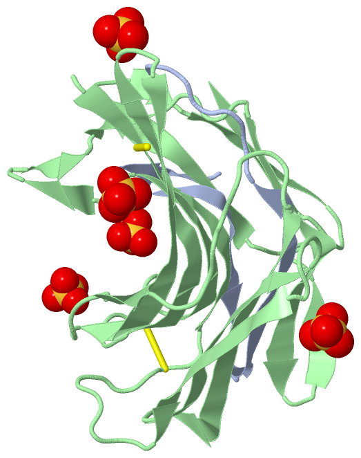
Asymmetric/Biological Unit (1, 5)
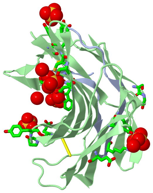
No.NameCountTypeFull Name
1SO45Ligand/IonSULFATE ION

(-) Sites  (5, 5)

Asymmetric Unit (5, 5)
No.NameEvidenceResiduesDescription
1AC1SOFTWARELYS A:19 , ALA A:31 , TYR B:5 , TYR B:31 , GLU B:68BINDING SITE FOR RESIDUE SO4 B 201
2AC2SOFTWARESER A:4 , ASN A:6 , TRP A:7 , TYR B:48BINDING SITE FOR RESIDUE SO4 A 202
3AC3SOFTWAREGLU B:19 , ASP B:28 , CYS B:30 , SER B:37 , PRO B:46 , HOH B:206BINDING SITE FOR RESIDUE SO4 B 203
4AC4SOFTWAREASP B:28 , PHE B:112 , HOH B:281 , HOH B:282BINDING SITE FOR RESIDUE SO4 B 204
5AC5SOFTWARELYS B:64 , GLU B:80 , SER B:164 , GLY B:165 , HOH B:254BINDING SITE FOR RESIDUE SO4 B 205

(-) SS Bonds  (2, 2)

Asymmetric/Biological Unit
No.Residues
1B:6 -B:30
2B:18 -B:101

(-) Cis Peptide Bonds  (1, 1)

Asymmetric/Biological Unit
No.Residues
1Tyr B:45 -Pro B:46

 Sequence-Structure Mapping

(-) SAPs(SNPs)/Variants  (0, 0)

(no "SAP(SNP)/Variant" information available for 1Y43)

(-) PROSITE Motifs  (0, 0)

(no "PROSITE Motif" information available for 1Y43)

(-) Exons   (0, 0)

(no "Exon" information available for 1Y43)

(-) Sequences/Alignments

Asymmetric/Biological Unit
   Reformat: Number of residues per line =  ('0' or empty: single-line sequence representation)
  Number of residues per labelling interval =   
  UniProt sequence: complete  aligned part    
   Show mapping: SCOP domains CATH domains Pfam domains Secondary structure (by author)
SAPs(SNPs) PROSITE motifs Exons
(details for a mapped element are shown in a popup box when the mouse pointer rests over it)
Chain A from PDB  Type:PROTEIN  Length:32
 aligned with PRTA_ASPNG | P24665 from UniProtKB/Swiss-Prot  Length:282

    Alignment length:32
                                    69        79        89  
           PRTA_ASPNG    60 EEYSSNWAGAVLIGDGYTKVTGEFTVPSVSAG  91
               SCOP domains d1y43.1 A:1-32,B:3-173           SCOP domains
               CATH domains -------------------------------- CATH domains
               Pfam domains -------------------------------- Pfam domains
         Sec.struct. author .eee..eeeeeee...eeeeeeeee....... Sec.struct. author
                 SAPs(SNPs) -------------------------------- SAPs(SNPs)
                    PROSITE -------------------------------- PROSITE
                 Transcript -------------------------------- Transcript
                 1y43 A   1 EEYSSNWAGAVLIGDGYTKVTGEFTVPSVSAG  32
                                    10        20        30  

Chain B from PDB  Type:PROTEIN  Length:171
 aligned with PRTA_ASPNG | P24665 from UniProtKB/Swiss-Prot  Length:282

    Alignment length:171
                                   121       131       141       151       161       171       181       191       201       211       221       231       241       251       261       271       281 
           PRTA_ASPNG   112 EEYCASAWVGIDGDTCETAILQTGVDFCYEDGQTSYDAWYEWYPDYAYDFSDITISEGDSIKVTVEATSKSSGSATVENLTTGQSVTHTFSGNVEGDLCETNAEWIVEDFESGDSLVAFADFGSVTFTNAEATSGGSTVGPSDATVMDIEQDGSVLTETSVSGDSVTVTYV 282
               SCOP domains d1y43.1 A:1-32,B:3-173 Aspergillopepsin II                                                                                                                                  SCOP domains
               CATH domains --------------------------------------------------------------------------------------------------------------------------------------------------------------------------- CATH domains
           Pfam domains (1) Peptidase_A4-1y43B01 B:3-173                                                                                                                                                Pfam domains (1)
           Pfam domains (2) Peptidase_A4-1y43B02 B:3-173                                                                                                                                                Pfam domains (2)
         Sec.struct. author .eeeeeeeeee........eeeeeeeeeee..eeeeeeeeeee....eee.........eeeeeeeeee..eeeeeeee....eeeeeee...........eeeeeee..ee..ee.ee....eeeeeeeeeee..eee.....eeeeeee..ee.eeeee....eeeee. Sec.struct. author
                 SAPs(SNPs) --------------------------------------------------------------------------------------------------------------------------------------------------------------------------- SAPs(SNPs)
                    PROSITE --------------------------------------------------------------------------------------------------------------------------------------------------------------------------- PROSITE
                 Transcript --------------------------------------------------------------------------------------------------------------------------------------------------------------------------- Transcript
                 1y43 B   3 EEYCASAWVGIDGDTCETAILQTGVDFCYEDGQTSYDAWYEWYPDYAYDFSDITISEGDSIKVTVEATSKSSGSATVENLTTGQSVTHTFSGNVEGDLCETNAEWIVEDFESGDSLVAFADFGSVTFTNAEATSGGSTVGPSDATVMDIEQDGSVLTETSVSGDSVTVTYV 173
                                    12        22        32        42        52        62        72        82        92       102       112       122       132       142       152       162       172 

   Legend:   → Mismatch (orange background)
  - → Gap (green background, '-', border residues have a numbering label)
    → Modified Residue (blue background, lower-case, 'x' indicates undefined single-letter code, labelled with number + name)
  x → Chemical Group (purple background, 'x', labelled with number + name, e.g. ACE or NH2)
  extra numbering lines below/above indicate numbering irregularities and modified residue names etc., number ends below/above '|'

 Classification and Annotation

(-) SCOP Domains  (1, 1)

Asymmetric/Biological Unit

(-) CATH Domains  (0, 0)

(no "CATH Domain" information available for 1Y43)

(-) Pfam Domains  (1, 2)

Asymmetric/Biological Unit

(-) Gene Ontology  (4, 4)

Asymmetric/Biological Unit(hide GO term definitions)
Chain A,B   (PRTA_ASPNG | P24665)
molecular function
    GO:0004190    aspartic-type endopeptidase activity    Catalysis of the hydrolysis of internal, alpha-peptide bonds in a polypeptide chain by a mechanism in which a water molecule bound by the side chains of aspartic residues at the active center acts as a nucleophile.
    GO:0016787    hydrolase activity    Catalysis of the hydrolysis of various bonds, e.g. C-O, C-N, C-C, phosphoric anhydride bonds, etc. Hydrolase is the systematic name for any enzyme of EC class 3.
    GO:0008233    peptidase activity    Catalysis of the hydrolysis of a peptide bond. A peptide bond is a covalent bond formed when the carbon atom from the carboxyl group of one amino acid shares electrons with the nitrogen atom from the amino group of a second amino acid.
biological process
    GO:0006508    proteolysis    The hydrolysis of proteins into smaller polypeptides and/or amino acids by cleavage of their peptide bonds.

 Visualization

(-) Interactive Views

Asymmetric/Biological Unit
  Complete Structure
    Jena3D(integrated viewing of ligand, site, SAP, PROSITE, SCOP information)
    WebMol | AstexViewer[tm]@PDBe
(Java Applets, require no local installation except for Java; loading may be slow)
    STRAP
(Java WebStart application, automatic local installation, requires Java; full application with system access!)
    RasMol
(require local installation)
    Molscript (VRML)
(requires installation of a VRML viewer; select preferred view via VRML and generate a mono or stereo PDF format file)
 
  Ligands, Modified Residues, Ions
    SO4  [ RasMol | Jena3D ]  +environment [ RasMol | Jena3D ]
 
  Sites
    AC1  [ RasMol ]  +environment [ RasMol ]
    AC2  [ RasMol ]  +environment [ RasMol ]
    AC3  [ RasMol ]  +environment [ RasMol ]
    AC4  [ RasMol ]  +environment [ RasMol ]
    AC5  [ RasMol ]  +environment [ RasMol ]
 
  Cis Peptide Bonds
    Tyr B:45 - Pro B:46   [ RasMol ]  
 

(-) Still Images

Jmol
  protein: cartoon or spacefill or dots and stick; nucleic acid: cartoon and stick; ligands: spacefill; active site: stick
Molscript
  protein, nucleic acid: cartoon; ligands: spacefill; active site: ball and stick

 Databases and Analysis Tools

(-) Databases

Access by PDB/NDB ID
  1y43
    Family and Domain InformationProDom | SYSTERS
    General Structural InformationGlycoscienceDB | MMDB | NDB | OCA | PDB | PDBe | PDBj | PDBsum | PDBWiki | PQS | PROTEOPEDIA
    Orientation in MembranesOPM
    Protein SurfaceSURFACE
    Secondary StructureDSSP (structure derived) | HSSP (homology derived)
    Structural GenomicsGeneCensus
    Structural NeighboursCE | VAST
    Structure ClassificationCATH | Dali | SCOP
    Validation and Original DataBMRB Data View | BMRB Restraints Grid | EDS | PROCHECK | RECOORD | WHAT_CHECK
 
Access by UniProt ID/Accession number
  PRTA_ASPNG | P24665
    Comparative Protein Structure ModelsModBase
    Genomic InformationEnsembl
    Protein-protein InteractionDIP
    Sequence, Family and Domain InformationInterPro | Pfam | SMART | UniProtKB/SwissProt
 
Access by Enzyme Classificator   (EC Number)
  3.4.23.19
    General Enzyme InformationBRENDA | EC-PDB | Enzyme | IntEnz
    PathwayKEGG | MetaCyc
 
Access by Disease Identifier   (MIM ID)
  (no 'MIM ID' available)
    Disease InformationOMIM
 
Access by GenAge ID
  (no 'GenAge ID' available)
    Age Related InformationGenAge

(-) Analysis Tools

Access by PDB/NDB ID
    Domain InformationXDom
    Interatomic Contacts of Structural UnitsCSU
    Ligand-protein ContactsLPC
    Protein CavitiescastP
    Sequence and Secondary StructurePDBCartoon
    Structure AlignmentSTRAP(Java WebStart application, automatic local installation, requires Java; full application with system access!)
    Structure and Sequence BrowserSTING
 
Access by UniProt ID/Accession number
  PRTA_ASPNG | P24665
    Protein Disorder PredictionDisEMBL | FoldIndex | GLOBPLOT (for more information see DisProt)

 Related Entries

(-) Entries Sharing at Least One Protein Chain (UniProt ID)

UniProtKB/Swiss-Prot
        PRTA_ASPNG | P246653trs

(-) Related Entries Specified in the PDB File

(no "Related Entries Specified in the PDB File" available for 1Y43)