Show PDB file:   
         Plain Text   HTML   (compressed file size)
QuickSearch:   
by PDB,NDB,UniProt,PROSITE Code or Search Term(s)  
(-)NMR Structure - model 1
(-)NMR Structure - all models
collapse expand < >
Image NMR Structure - model 1
NMR Structure - model 1  (Jmol Viewer)
Image NMR Structure - all models
NMR Structure - all models  (Jmol Viewer)

(-) Description

Title :  STRUCTURE OF THE C-TERMINAL DOMAIN FROM TRYPANOSOMA BRUCEI VARIANT SURFACE GLYCOPROTEIN MITAT1.2
 
Authors :  A. Chattopadhyay, N. G. Jones, D. Nietlispach, P. R. Nielsen, H. P. Voor H. R. Mott, M. Carrington
Date :  25 Oct 04  (Deposition) - 30 Nov 04  (Release) - 13 Jul 11  (Revision)
Method :  SOLUTION NMR
Resolution :  NOT APPLICABLE
Chains :  NMR Structure  :  A  (60x)
NMR Structure *:  A  (1x)
Keywords :  Cysteine Knot, Immune System, Membrane Protein (Keyword Search: [Gene Ontology, PubMed, Web (Google))
 
Reference :  A. Chattopadhyay, N. G. Jones, D. Nietlispach, P. R. Nielsen, H. P. Voorheis, H. R. Mott, M. Carrington
Structure Of The C-Terminal Domain From Trypanosoma Brucei Variant Surface Glycoprotein Mitat1. 2
J. Biol. Chem. V. 280 7228 2004
PubMed-ID: 15557330  |  Reference-DOI: 10.1074/JBC.M410787200

(-) Compounds

Molecule 1 - VARIANT SURFACE GLYCOPROTEIN MITAT 1.2
    ChainsA
    EngineeredYES
    Expression SystemESCHERICHIA COLI
    Expression System PlasmidPET15B
    Expression System StrainBL21 TRXB
    Expression System Taxid562
    Expression System Vector TypePLASMID
    Organism ScientificTRYPANOSOMA
    Organism Taxid5702
    StrainBRUCEI
    SynonymVSG 221, MITAT1.2 C-TERMINAL DOMAIN

 Structural Features

(-) Chains, Units

  1
NMR Structure (60x)A
NMR Structure * (1x)A

Summary Information (see also Sequences/Alignments below)

(-) Ligands, Modified Residues, Ions  (0, 0)

(no "Ligand,Modified Residues,Ions" information available for 1XU6)

(-) Sites  (0, 0)

(no "Site" information available for 1XU6)
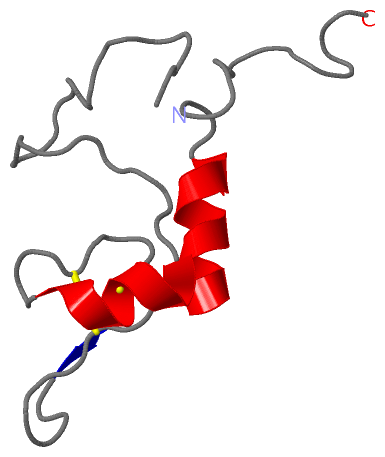
(-) SS Bonds  (2, 2)

NMR Structure
No.Residues
1A:381 -A:393
2A:389 -A:404

(-) Cis Peptide Bonds  (0, 0)

(no "Cis Peptide Bond" information available for 1XU6)

 Sequence-Structure Mapping

(-) SAPs(SNPs)/Variants  (0, 0)

(no "SAP(SNP)/Variant" information available for 1XU6)

(-) PROSITE Motifs  (0, 0)

(no "PROSITE Motif" information available for 1XU6)

(-) Exons   (0, 0)

(no "Exon" information available for 1XU6)

(-) Sequences/Alignments

NMR Structure
   Reformat: Number of residues per line =  ('0' or empty: single-line sequence representation)
  Number of residues per labelling interval =   
  UniProt sequence: complete  aligned part    
   Show mapping: SCOP domains CATH domains Pfam domains Secondary structure (by author)
SAPs(SNPs) PROSITE motifs Exons
(details for a mapped element are shown in a popup box when the mouse pointer rests over it)
Chain A from PDB  Type:PROTEIN  Length:80
 aligned with VSM2_TRYBB | P26332 from UniProtKB/Swiss-Prot  Length:476

    Alignment length:95
                                   374       384       394       404       414       424       434       444       454     
           VSM2_TRYBB   365 GNAKLTTILAYYRMETAGKFEVLTQKHKPAESQQQAAETEGSCNKKDQNECKSPCKWHNDAENKKCTLDKEEAKKVADETAKDGKTGNTNTTGSS 459
               SCOP domains d               1xu6a_ A: Variant surface glycoprotein MITAT 1.2, VSG 221, C-terminal domain    SCOP domains
               CATH domains 1               xu6A00 A:354-433 Variant surface glycoprotein mitat 1.2                         CATH domains
               Pfam domains ----------------------------------------------------------------------------------------------- Pfam domains
         Sec.struct. author .---------------......................hhhhhhhh..........ee..........hhhhhhhhhh................. Sec.struct. author
                 SAPs(SNPs) ----------------------------------------------------------------------------------------------- SAPs(SNPs)
                    PROSITE ----------------------------------------------------------------------------------------------- PROSITE
                 Transcript ----------------------------------------------------------------------------------------------- Transcript
                 1xu6 A 354 G---------------SHMLEVLTQKHKPAESQQQAAETEGSCNKKDQNECKSPCKWHNDAENKKCTLDKEEAKKVADETAKDGKTGNTNTTGSS 433
                            |        -      |358       368       378       388       398       408       418       428     
                            |             355                                                                              
                          354                                                                                              

   Legend:   → Mismatch (orange background)
  - → Gap (green background, '-', border residues have a numbering label)
    → Modified Residue (blue background, lower-case, 'x' indicates undefined single-letter code, labelled with number + name)
  x → Chemical Group (purple background, 'x', labelled with number + name, e.g. ACE or NH2)
  extra numbering lines below/above indicate numbering irregularities and modified residue names etc., number ends below/above '|'

 Classification and Annotation

(-) SCOP Domains  (1, 1)

NMR Structure

(-) CATH Domains  (1, 1)

NMR Structure

(-) Pfam Domains  (0, 0)

(no "Pfam Domain" information available for 1XU6)

(-) Gene Ontology  (4, 4)

NMR Structure(hide GO term definitions)
Chain A   (VSM2_TRYBB | P26332)
biological process
    GO:0020012    evasion or tolerance of host immune response    Any process, either active or passive, by which an organism avoids the effects of the host organism's immune response. The host is defined as the larger of the organisms involved in a symbiotic interaction.
cellular component
    GO:0031225    anchored component of membrane    The component of a membrane consisting of the gene products that are tethered to the membrane only by a covalently attached anchor, such as a lipid group that is embedded in the membrane. Gene products with peptide sequences that are embedded in the membrane are excluded from this grouping.
    GO:0016020    membrane    A lipid bilayer along with all the proteins and protein complexes embedded in it an attached to it.
    GO:0005886    plasma membrane    The membrane surrounding a cell that separates the cell from its external environment. It consists of a phospholipid bilayer and associated proteins.

 Visualization

(-) Interactive Views

NMR Structure
  Complete Structure
    Jena3D(integrated viewing of ligand, site, SAP, PROSITE, SCOP information)
    WebMol | AstexViewer[tm]@PDBe
(Java Applets, require no local installation except for Java; loading may be slow)
    STRAP
(Java WebStart application, automatic local installation, requires Java; full application with system access!)
    RasMol
(require local installation)
    Molscript (VRML)
(requires installation of a VRML viewer; select preferred view via VRML and generate a mono or stereo PDF format file)
 
  Ligands, Modified Residues, Ions
(no "Ligands, Modified Residues, Ions" information available for 1xu6)
 
  Sites
(no "Sites" information available for 1xu6)
 
  Cis Peptide Bonds
(no "Cis Peptide Bonds" information available for 1xu6)
 

(-) Still Images

Jmol
  protein: cartoon or spacefill or dots and stick; nucleic acid: cartoon and stick; ligands: spacefill; active site: stick
Molscript
  protein, nucleic acid: cartoon; ligands: spacefill; active site: ball and stick

 Databases and Analysis Tools

(-) Databases

Access by PDB/NDB ID
  1xu6
    Family and Domain InformationProDom | SYSTERS
    General Structural InformationGlycoscienceDB | MMDB | NDB | OCA | PDB | PDBe | PDBj | PDBsum | PDBWiki | PQS | PROTEOPEDIA
    Orientation in MembranesOPM
    Protein SurfaceSURFACE
    Secondary StructureDSSP (structure derived) | HSSP (homology derived)
    Structural GenomicsGeneCensus
    Structural NeighboursCE | VAST
    Structure ClassificationCATH | Dali | SCOP
    Validation and Original DataBMRB Data View | BMRB Restraints Grid | EDS | PROCHECK | RECOORD | WHAT_CHECK
 
Access by UniProt ID/Accession number
  VSM2_TRYBB | P26332
    Comparative Protein Structure ModelsModBase
    Genomic InformationEnsembl
    Protein-protein InteractionDIP
    Sequence, Family and Domain InformationInterPro | Pfam | SMART | UniProtKB/SwissProt
 
Access by Enzyme Classificator   (EC Number)
  (no 'Enzyme Classificator' available)
    General Enzyme InformationBRENDA | EC-PDB | Enzyme | IntEnz
    PathwayKEGG | MetaCyc
 
Access by Disease Identifier   (MIM ID)
  (no 'MIM ID' available)
    Disease InformationOMIM
 
Access by GenAge ID
  (no 'GenAge ID' available)
    Age Related InformationGenAge

(-) Analysis Tools

Access by PDB/NDB ID
    Domain InformationXDom
    Interatomic Contacts of Structural UnitsCSU
    Ligand-protein ContactsLPC
    Protein CavitiescastP
    Sequence and Secondary StructurePDBCartoon
    Structure AlignmentSTRAP(Java WebStart application, automatic local installation, requires Java; full application with system access!)
    Structure and Sequence BrowserSTING
 
Access by UniProt ID/Accession number
  VSM2_TRYBB | P26332
    Protein Disorder PredictionDisEMBL | FoldIndex | GLOBPLOT (for more information see DisProt)

 Related Entries

(-) Entries Sharing at Least One Protein Chain (UniProt ID)

UniProtKB/Swiss-Prot
        VSM2_TRYBB | P263321vsg

(-) Related Entries Specified in the PDB File

(no "Related Entries Specified in the PDB File" available for 1XU6)