|
|
|
|
 Description
|
|
Compounds
|
||||||||||||||||||||||||||||||||
Chains, Units
Summary Information (see also Sequences/Alignments below) |
Ligands, Modified Residues, Ions (0, 0)| (no "Ligand,Modified Residues,Ions" information available for 1XG7) |
Sites (0, 0)| (no "Site" information available for 1XG7) |
SS Bonds (0, 0)| (no "SS Bond" information available for 1XG7) |
Cis Peptide Bonds (0, 0)| (no "Cis Peptide Bond" information available for 1XG7) |
SAPs(SNPs)/Variants (0, 0)| (no "SAP(SNP)/Variant" information available for 1XG7) |
PROSITE Motifs (0, 0)| (no "PROSITE Motif" information available for 1XG7) |
Exons (0, 0)| (no "Exon" information available for 1XG7) |
Sequences/Alignments
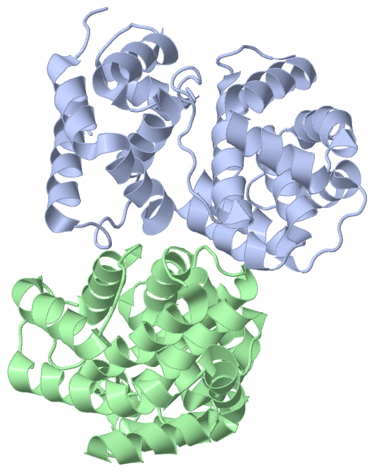
Asymmetric/Biological UnitChain A from PDB Type:PROTEIN Length:245 aligned with AGOG_PYRFU | Q8U2D5 from UniProtKB/Swiss-Prot Length:242 Alignment length:245 1 | 7 17 27 37 47 57 67 77 87 97 107 117 127 137 147 157 167 177 187 197 207 217 227 237 AGOG_PYRFU - ---MRELVEIIKGIGIEGAKEVEEKVDRQFYALQYLFRHQDPEMFIKLVIANSLVSYQLTGRGEDWWWEFARYFSGREVDSIWKAYGEFLPKSKNNRRLIEAKLNRIRKVEGFLSTLTLKDLEGYYKNMKMLWKALIKIMGSREDSKTIVFTVKMFGYASRIAFSRFIPYPMEIPIPEDLRIKSVTSKLTQEKPTKFWMKIGQESGVPPLHIDSLIWPLLGNADLTPLDIELRNKLMKLTELLGL 242 SCOP domains d1xg7a_ A: Hypothetical protein PF0904 SCOP domains CATH domains 1xg7A01 A:-2-23,A:167-242 1xg7A02 A:24-166 Hypothetical protein; domain 2 1xg7A01 A:-2-23,A:167-242 CATH domains Pfam domains ----------------------------------------------------------------------------------------------------------------------------------------------------------------------------------------------------------------------------------------------------- Pfam domains SAPs(SNPs) ----------------------------------------------------------------------------------------------------------------------------------------------------------------------------------------------------------------------------------------------------- SAPs(SNPs) PROSITE ----------------------------------------------------------------------------------------------------------------------------------------------------------------------------------------------------------------------------------------------------- PROSITE Transcript ----------------------------------------------------------------------------------------------------------------------------------------------------------------------------------------------------------------------------------------------------- Transcript 1xg7 A -2 HHGSRELVEIIKGIGIEGAKEVEEKVDRQFYALQYLFRHQDPEMFIKLVIANSLVSYQLTGRGEDWWWEFARYFSGREVDSIWKAYGEFLPKSKNNRRLIEAKLNRIRKVEGFLSTLTLKDLEGYYKNMKMLWKALIKIMGSREDSKTIVFTVKMFGYASRIAFSRFIPYPMEIPIPEDLRIKSVTSKLTQEKPTKFWMKIGQESGVPPLHIDSLIWPLLGNADLTPLDIELRNKLMKLTELLGL 242 7 17 27 37 47 57 67 77 87 97 107 117 127 137 147 157 167 177 187 197 207 217 227 237 Chain B from PDB Type:PROTEIN Length:239 aligned with AGOG_PYRFU | Q8U2D5 from UniProtKB/Swiss-Prot Length:242 Alignment length:239 12 22 32 42 52 62 72 82 92 102 112 122 132 142 152 162 172 182 192 202 212 222 232 AGOG_PYRFU 3 ELVEIIKGIGIEGAKEVEEKVDRQFYALQYLFRHQDPEMFIKLVIANSLVSYQLTGRGEDWWWEFARYFSGREVDSIWKAYGEFLPKSKNNRRLIEAKLNRIRKVEGFLSTLTLKDLEGYYKNMKMLWKALIKIMGSREDSKTIVFTVKMFGYASRIAFSRFIPYPMEIPIPEDLRIKSVTSKLTQEKPTKFWMKIGQESGVPPLHIDSLIWPLLGNADLTPLDIELRNKLMKLTELLG 241 SCOP domains d1xg7b_ B: Hypothetical protein PF0904 SCOP domains CATH domains 1xg7B01 1xg7B02 B:24-166 Hypothetical protein; domain 2 1xg7B01 B:3-23,B:167-241 CATH domains Pfam domains (1) -DUF1886-1xg7B01 B:4-238 --- Pfam domains (1) Pfam domains (2) -DUF1886-1xg7B02 B:4-238 --- Pfam domains (2) SAPs(SNPs) ----------------------------------------------------------------------------------------------------------------------------------------------------------------------------------------------------------------------------------------------- SAPs(SNPs) PROSITE ----------------------------------------------------------------------------------------------------------------------------------------------------------------------------------------------------------------------------------------------- PROSITE Transcript ----------------------------------------------------------------------------------------------------------------------------------------------------------------------------------------------------------------------------------------------- Transcript 1xg7 B 3 ELVEIIKGIGIEGAKEVEEKVDRQFYALQYLFRHQDPEMFIKLVIANSLVSYQLTGRGEDWWWEFARYFSGREVDSIWKAYGEFLPKSKNNRRLIEAKLNRIRKVEGFLSTLTLKDLEGYYKNMKMLWKALIKIMGSREDSKTIVFTVKMFGYASRIAFSRFIPYPMEIPIPEDLRIKSVTSKLTQEKPTKFWMKIGQESGVPPLHIDSLIWPLLGNADLTPLDIELRNKLMKLTELLG 241 12 22 32 42 52 62 72 82 92 102 112 122 132 142 152 162 172 182 192 202 212 222 232
|
||||||||||||||||||||
SCOP Domains (1, 2)
Asymmetric/Biological Unit
 |
CATH Domains (2, 4)
Asymmetric/Biological Unit
|
Pfam Domains (1, 2)
Asymmetric/Biological Unit
|
Gene Ontology (9, 9)|
Asymmetric/Biological Unit(hide GO term definitions) Chain A,B (AGOG_PYRFU | Q8U2D5)
|
||||||||||||||||||||||||||||||||||||||||||||||||||||||||||||||||||
Interactive Views
|
||||||||||||||||||||||||||||||||||||||||||||||||||||||||||||||||||||||||||||||||||||||||||||||||||||||||||||||||||||
Still Images
|
||||||||||||||||
Databases
|
||||||||||||||||||||||||||||||||||||||||||||||||||||||||||||||||||||||||||||||||||||||||||||||||||||||||||||||||||||||||||||||||||||||||||||||||||||||||||||||||
Analysis Tools
|
|||||||||||||||||||||||||||||||||||||||||||||||||||||||||||||
Entries Sharing at Least One Protein Chain (UniProt ID)
Related Entries Specified in the PDB File
|
|