|
|
|
|
 Description
|
|
Compounds
|
||||||||||||||||||||||||||||||||||||||||||||
Chains, Units
Summary Information (see also Sequences/Alignments below) |
Ligands, Modified Residues, Ions (0, 0)| (no "Ligand,Modified Residues,Ions" information available for 1WYJ) |
Sites (0, 0)| (no "Site" information available for 1WYJ) |
SS Bonds (1, 1)
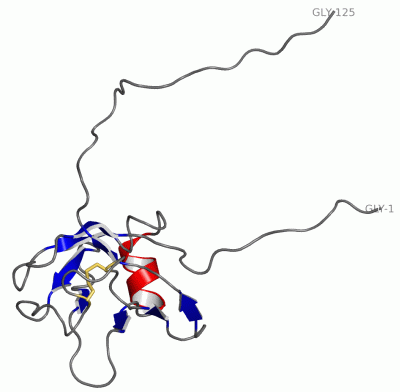
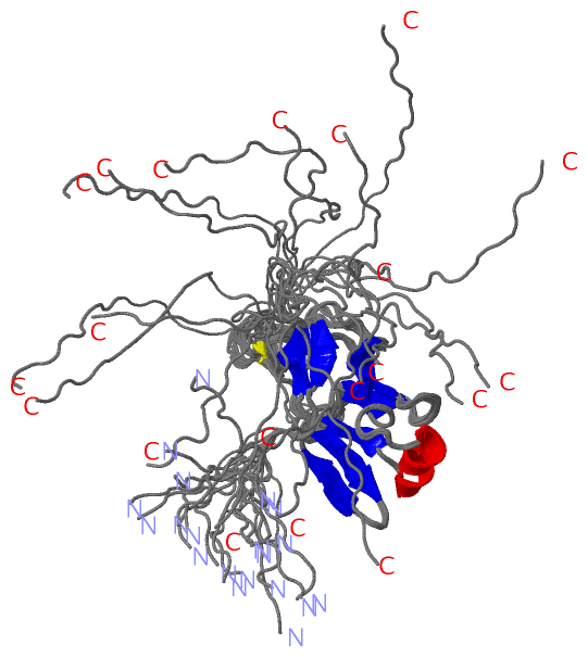
NMR Structure
|
||||||||
Cis Peptide Bonds (2, 40)
NMR Structure
|
|||||||||||||||
SAPs(SNPs)/Variants (0, 0)| (no "SAP(SNP)/Variant" information available for 1WYJ) |
PROSITE Motifs (2, 3)
NMR Structure (2, 3)
|
||||||||||||||||||||||||||||||||
Exons (0, 0)| (no "Exon" information available for 1WYJ) |
Sequences/Alignments
NMR StructureChain A from PDB Type:PROTEIN Length:125 aligned with PCDBE_MOUSE | Q6PB90 from UniProtKB/Swiss-Prot Length:796 Alignment length:223 11 21 31 41 51 61 71 81 91 101 111 121 131 141 151 161 171 181 191 201 211 221 PCDBE_MOUSE 2 ETSLHKAPQKRQVTAIIFLLLLWEAGSATITYSVLEETDRGSLVGNLAKDLGLSLRELITRGAQILSKGNKQLLQLEQKSGNLLLKEKLDREELCGSTNPCILHFQVLLKSPVQFIQGEIQLQDVNDHAPEFMEDEILLKILESSLPGAVFPLKIAQDLDVGSNTVQNYTISTNAHFHLLTRNHSDGRKYPELVLDKALDREEQAQIRLTLTAMDSGSPPKTG 224 SCOP domains ------------------------------------------------------------------------------------------------------------------------------------------------------------------------------------------------------------------------------- SCOP domains CATH domains ------------------------------------------------------------------------------------------------------------------------------------------------------------------------------------------------------------------------------- CATH domains Pfam domains ---------------------------Cadherin_2-1wyjA01 A:11-94 ---------------------------------------------------------------------------------------------------------------- Pfam domains SAPs(SNPs) ------------------------------------------------------------------------------------------------------------------------------------------------------------------------------------------------------------------------------- SAPs(SNPs) PROSITE (1) --------------------------CADHERIN_2 PDB: A:10-115 UniProt: 28-133 CADHERIN_2 PDB: A:116-125 UniProt: 134-242 PROSITE (1) PROSITE (2) -----------------------------------------------------------------------------------------------------------------------CADHERIN_1 --------------------------------------------------------------------------------------------- PROSITE (2) Transcript ------------------------------------------------------------------------------------------------------------------------------------------------------------------------------------------------------------------------------- Transcript 1wyj A 1 GSSGSSG-----------------AGSATITYSVLEETDRGSLVGNLAKDLGLSLRELITRGAQILSKGNKQLLQLEQKSGNLLLKEKLDREELCGSTNPCILHFQVLLKSPVQFIQGEIQLQDVNDHAPEFMEDE-------------------------------------------------------------------------------SG--PSSG 125 | - - | 13 23 33 43 53 63 73 83 93 103 113 | - - - - - - - - ||122 7 8 119 120| | 121 | 122
|
||||||||||||||||||||
SCOP Domains (0, 0)| (no "SCOP Domain" information available for 1WYJ) |
CATH Domains (0, 0)| (no "CATH Domain" information available for 1WYJ) |
Pfam Domains (1, 1)
NMR Structure

|
Gene Ontology (6, 6)|
NMR Structure(hide GO term definitions) Chain A (PCDBE_MOUSE | Q6PB90)
|
||||||||||||||||||||||||||||||||||||||||||||||||||||||
Interactive Views
|
||||||||||||||||||||||||||||||||||||||||||||||||||||||||||||||||||||||||||||||||||||||||||||||||||||||||||||||||||||||||||||
Still Images
|
||||||||||||||||
Databases
|
||||||||||||||||||||||||||||||||||||||||||||||||||||||||||||||||||||||||||||||||||||||||||||||||||||||||||||||||||||||||||||||||||||||||||||||||||||||||||||||||
Analysis Tools
|
|||||||||||||||||||||||||||||||||||||||||||||||||||||||||||||
Entries Sharing at Least One Protein Chain (UniProt ID)
Related Entries Specified in the PDB File
|
|