|
|
|
|
 Description
|
|
Compounds
|
||||||||||||||||||||||||||||||||||||||||||||
Chains, Units
Summary Information (see also Sequences/Alignments below) |
Ligands, Modified Residues, Ions (0, 0)| (no "Ligand,Modified Residues,Ions" information available for 1WRY) |
Sites (0, 0)| (no "Site" information available for 1WRY) |
SS Bonds (0, 0)| (no "SS Bond" information available for 1WRY) |
Cis Peptide Bonds (1, 20)
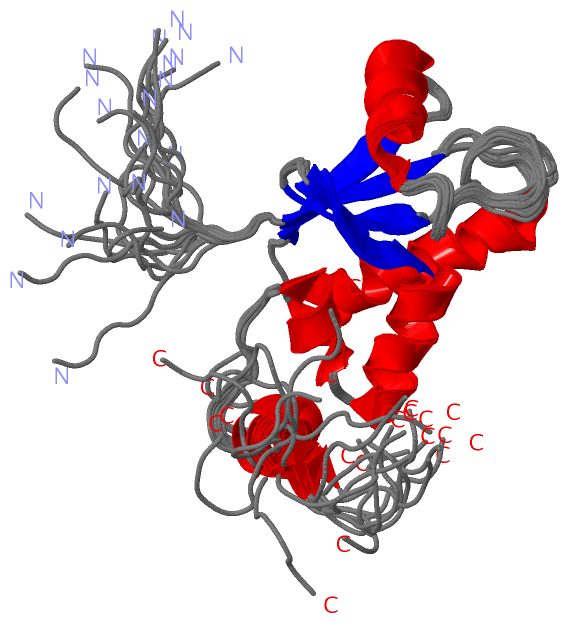
NMR Structure
|
||||||||||
SAPs(SNPs)/Variants (0, 0)| (no "SAP(SNP)/Variant" information available for 1WRY) |
PROSITE Motifs (0, 0)| (no "PROSITE Motif" information available for 1WRY) |
Exons (4, 4)
NMR Structure (4, 4)
|
||||||||||||||||||||||||||||||||||||||||||||||||||||||||||||||||||||||||
Sequences/Alignments
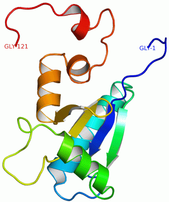
NMR StructureChain A from PDB Type:PROTEIN Length:121 aligned with SH3L1_HUMAN | O75368 from UniProtKB/Swiss-Prot Length:114 Alignment length:121 1 | 3 13 23 33 43 53 63 73 83 93 103 113 SH3L1_HUMAN - -------MVIRVYIASSSGSTAIKKKQQDVLGFLEANKIGFEEKDIAANEENRKWMRENVPENSRPATGYPLPPQIFNESQYRGDYDAFFEARENNAVYAFLGLTAPPGSKEAEVQAKQQA 114 SCOP domains d1wrya_ A: automated matches SCOP domains CATH domains 1wryA00 A:1-121 Glutaredoxin CATH domains Pfam domains -------SH3BGR-1wryA01 A:8-105 ---------------- Pfam domains SAPs(SNPs) ------------------------------------------------------------------------------------------------------------------------- SAPs(SNPs) PROSITE ------------------------------------------------------------------------------------------------------------------------- PROSITE Transcript 1 -------Exon 1.1a Exon 1.5 PDB: A:23-84 UniProt: 16-77 Exon 1.6 PDB: A:85-111 Exon 1.7d Transcript 1 1wry A 1 GSSGSSGMVIRVYIASSSGSTAIKKKQQDVLGFLEANKIGFEEKDIAANEENRKWMRENVPENSRPATGYPLPPQIFNESQYRGDYDAFFEARENNAVYAFLGLTAPPGSKEAEVSGPSSG 121 10 20 30 40 50 60 70 80 90 100 110 120
|
||||||||||||||||||||
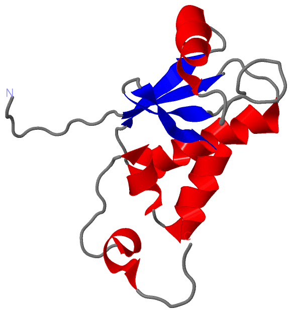
SCOP Domains (1, 1)
NMR Structure
 |
CATH Domains (1, 1)
NMR Structure
|
Pfam Domains (1, 1)| NMR Structure |
Gene Ontology (7, 7)|
NMR Structure(hide GO term definitions) Chain A (SH3L1_HUMAN | O75368)
|
||||||||||||||||||||||||||||||||||||||||||||||||||||||||||||
Interactive Views
|
|||||||||||||||||||||||||||||||||||||||||||||||||||||||||||||||||||||||||||||||||||||||||||||||||||||||||||||||||||||
Still Images
|
||||||||||||||||
Databases
|
||||||||||||||||||||||||||||||||||||||||||||||||||||||||||||||||||||||||||||||||||||||||||||||||||||||||||||||||||||||||||||||||||||||||||||||||||||||||||||||||
Analysis Tools
|
|||||||||||||||||||||||||||||||||||||||||||||||||||||||||||||
Entries Sharing at Least One Protein Chain (UniProt ID)
Related Entries Specified in the PDB File
|
|