Show PDB file:   
         Plain Text   HTML   (compressed file size)
QuickSearch:   
by PDB,NDB,UniProt,PROSITE Code or Search Term(s)  
(-)Asym./Biol. Unit
(-)Asym./Biol. Unit - sites
collapse expand < >
Image Asym./Biol. Unit
Asym./Biol. Unit  (Jmol Viewer)
Image Asym./Biol. Unit - sites
Asym./Biol. Unit - sites  (Jmol Viewer)

(-) Description

Title :  CRYSTAL STRUCTURE OF H2-PROTEINASE
 
Authors :  T. Kumasaka, M. Yamamoto, H. Moriyama, N. Tanaka, M. Sato, Y. Katsube, Y. Yamakawa, T. Omori-Satoh, S. Iwanaga, T. Ueki
Date :  04 Aug 04  (Deposition) - 17 Aug 04  (Release) - 24 Feb 09  (Revision)
Method :  X-RAY DIFFRACTION
Resolution :  2.20
Chains :  Asym./Biol. Unit :  A
Keywords :  Metalloprotease, Zinc, Metzincin, Snake Venom, Hydrolase (Keyword Search: [Gene Ontology, PubMed, Web (Google))
 
Reference :  T. Kumasaka, M. Yamamoto, H. Moriyama, N. Tanaka, M. Sato, Y. Katsube, Y. Yamakawa, T. Omori-Satoh, S. Iwanaga, T. Ueki
Crystal Structure Of H2-Proteinase From The Venom Of Trimeresurus Flavoviridis
J. Biochem. (Tokyo) V. 119 49 1996
PubMed-ID: 8907175
(for further references see the PDB file header)

(-) Compounds

Molecule 1 - TRIMERELYSIN II
    ChainsA
    EC Number3.4.24.53
    Organism ScientificTRIMERESURUS FLAVOVIRIDIS
    Organism Taxid88087
    SynonymH2-PROTEINASE, H2 METALLOPROTEINASE

 Structural Features

(-) Chains, Units

  1
Asymmetric/Biological Unit A

Summary Information (see also Sequences/Alignments below)

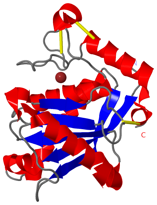
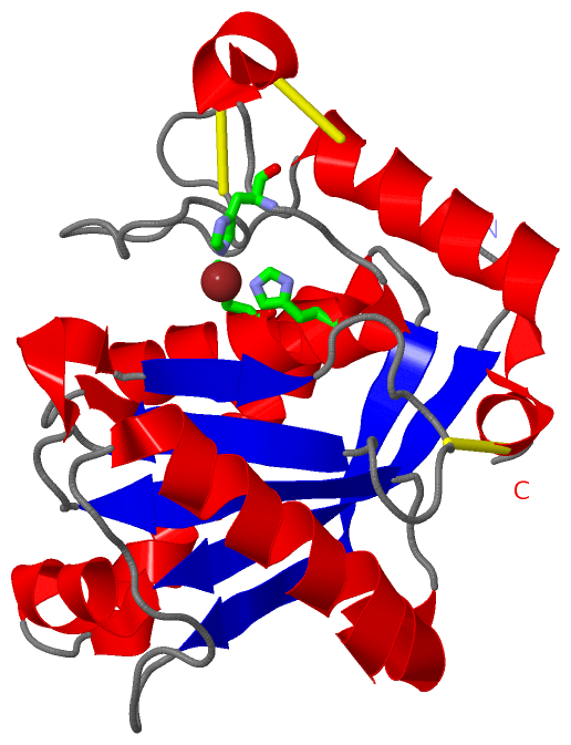
(-) Ligands, Modified Residues, Ions  (1, 1)

Asymmetric/Biological Unit (1, 1)
No.NameCountTypeFull Name
1ZN1Ligand/IonZINC ION

(-) Sites  (1, 1)

Asymmetric Unit (1, 1)
No.NameEvidenceResiduesDescription
1AC1SOFTWAREHIS A:142 , HIS A:146 , HIS A:152 , HOH A:1011BINDING SITE FOR RESIDUE ZN A 999

(-) SS Bonds  (3, 3)

Asymmetric/Biological Unit
No.Residues
1A:117 -A:196
2A:158 -A:180
3A:160 -A:163

(-) Cis Peptide Bonds  (0, 0)

(no "Cis Peptide Bond" information available for 1WNI)

 Sequence-Structure Mapping

(-) SAPs(SNPs)/Variants  (0, 0)

(no "SAP(SNP)/Variant" information available for 1WNI)

(-) PROSITE Motifs  (2, 2)

Asymmetric/Biological Unit (2, 2)
 PROSITEUniProtKBPDB
No.IDACDescriptionIDLocationCountLocation
1ADAM_MEPROPS50215 ADAM type metalloprotease domain profile.VM1T2_PROFL6-201  1A:6-200
2ZINC_PROTEASEPS00142 Neutral zinc metallopeptidases, zinc-binding region signature.VM1T2_PROFL139-148  1A:139-148

(-) Exons   (0, 0)

(no "Exon" information available for 1WNI)

(-) Sequences/Alignments

Asymmetric/Biological Unit
   Reformat: Number of residues per line =  ('0' or empty: single-line sequence representation)
  Number of residues per labelling interval =   
  UniProt sequence: complete  aligned part    
   Show mapping: SCOP domains CATH domains Pfam domains Secondary structure (by author)
SAPs(SNPs) PROSITE motifs Exons
(details for a mapped element are shown in a popup box when the mouse pointer rests over it)
Chain A from PDB  Type:PROTEIN  Length:198
 aligned with VM1T2_PROFL | P20165 from UniProtKB/Swiss-Prot  Length:201

    Alignment length:198
                                    12        22        32        42        52        62        72        82        92       102       112       122       132       142       152       162       172       182       192        
          VM1T2_PROFL     3 FPQRYIELAIVVDHGMYKKYNQNSDKIKVRVHQMVNHINEMYRPLNIAISLNRLQIWSKKDLITVKSASNVTLESFGNWRETVLLKQQNNDCAHLLTATNLNDNTIGLAYKKGMCNPKLSVGLVQDYSPNVFMVAVTMTHELGHNLGMEHDDKDKCKCEACIMSDVISDKPSKLFSDCSKNDYQTFLTKYNPQCILNA 200
               SCOP domains d1wnia_ A: Snake venom metalloprotease                                                                                                                                                                 SCOP domains
               CATH domains 1wniA00 A:3-200 Collagenase (Catalytic Domain)                                                                                                                                                         CATH domains
               Pfam domains ------------------------------------------------------------------------------------------------------------------------------------------------------------------------------------------------------ Pfam domains
         Sec.struct. author ...eeeeeeeeehhhhhhhh..hhhhhhhhhhhhhhhhhhhhh...eeeeeeeeee...........hhhhhhhhhhhhhhhhhhhhh...eeeeee...hhhhh.eee...........eeeee....hhhhhhhhhhhhhhhhh.....hhhhhh..................hhhhhhhhhhhhhhhhhhhhh.. Sec.struct. author
                 SAPs(SNPs) ------------------------------------------------------------------------------------------------------------------------------------------------------------------------------------------------------ SAPs(SNPs)
                PROSITE (1) ---ADAM_MEPRO  PDB: A:6-200 UniProt: 6-201                                                                                                                                                             PROSITE (1)
                PROSITE (2) ----------------------------------------------------------------------------------------------------------------------------------------ZINC_PROTE---------------------------------------------------- PROSITE (2)
                 Transcript ------------------------------------------------------------------------------------------------------------------------------------------------------------------------------------------------------ Transcript
                 1wni A   3 FPQRYIELAIVVDHGMYKKYNQNSDKIKVRVHQMVNHINEMYRPLNIAISLNRLQIWSKKDLITVKSASNVTLESFGNWRETVLLKQQNNDCAHLLTATNLNDNTIGLAYKKGMCNPKLSVGLVQDYSPNVFMVAVTMTHELGHNLGMEHDDKDKCKCEACIMSDVISDKPSKLFSDCSKNDYQTFLTKYNPQCILNA 200
                                    12        22        32        42        52        62        72        82        92       102       112       122       132       142       152       162       172       182       192        

   Legend:   → Mismatch (orange background)
  - → Gap (green background, '-', border residues have a numbering label)
    → Modified Residue (blue background, lower-case, 'x' indicates undefined single-letter code, labelled with number + name)
  x → Chemical Group (purple background, 'x', labelled with number + name, e.g. ACE or NH2)
  extra numbering lines below/above indicate numbering irregularities and modified residue names etc., number ends below/above '|'

 Classification and Annotation

(-) SCOP Domains  (1, 1)

Asymmetric/Biological Unit

(-) CATH Domains  (1, 1)

Asymmetric/Biological Unit
(-)
Class: Alpha Beta (26913)

(-) Pfam Domains  (0, 0)

(no "Pfam Domain" information available for 1WNI)

(-) Gene Ontology  (7, 7)

Asymmetric/Biological Unit(hide GO term definitions)
Chain A   (VM1T2_PROFL | P20165)
molecular function
    GO:0016787    hydrolase activity    Catalysis of the hydrolysis of various bonds, e.g. C-O, C-N, C-C, phosphoric anhydride bonds, etc. Hydrolase is the systematic name for any enzyme of EC class 3.
    GO:0046872    metal ion binding    Interacting selectively and non-covalently with any metal ion.
    GO:0004222    metalloendopeptidase activity    Catalysis of the hydrolysis of internal, alpha-peptide bonds in a polypeptide chain by a mechanism in which water acts as a nucleophile, one or two metal ions hold the water molecule in place, and charged amino acid side chains are ligands for the metal ions.
    GO:0008237    metallopeptidase activity    Catalysis of the hydrolysis of peptide bonds by a mechanism in which water acts as a nucleophile, one or two metal ions hold the water molecule in place, and charged amino acid side chains are ligands for the metal ions.
    GO:0008233    peptidase activity    Catalysis of the hydrolysis of a peptide bond. A peptide bond is a covalent bond formed when the carbon atom from the carboxyl group of one amino acid shares electrons with the nitrogen atom from the amino group of a second amino acid.
biological process
    GO:0006508    proteolysis    The hydrolysis of proteins into smaller polypeptides and/or amino acids by cleavage of their peptide bonds.
cellular component
    GO:0005576    extracellular region    The space external to the outermost structure of a cell. For cells without external protective or external encapsulating structures this refers to space outside of the plasma membrane. This term covers the host cell environment outside an intracellular parasite.

 Visualization

(-) Interactive Views

Asymmetric/Biological Unit
  Complete Structure
    Jena3D(integrated viewing of ligand, site, SAP, PROSITE, SCOP information)
    WebMol | AstexViewer[tm]@PDBe
(Java Applets, require no local installation except for Java; loading may be slow)
    STRAP
(Java WebStart application, automatic local installation, requires Java; full application with system access!)
    RasMol
(require local installation)
    Molscript (VRML)
(requires installation of a VRML viewer; select preferred view via VRML and generate a mono or stereo PDF format file)
 
  Ligands, Modified Residues, Ions
    ZN  [ RasMol | Jena3D ]  +environment [ RasMol | Jena3D ]
 
  Sites
    AC1  [ RasMol ]  +environment [ RasMol ]
 
  Cis Peptide Bonds
(no "Cis Peptide Bonds" information available for 1wni)
 

(-) Still Images

Jmol
  protein: cartoon or spacefill or dots and stick; nucleic acid: cartoon and stick; ligands: spacefill; active site: stick
Molscript
  protein, nucleic acid: cartoon; ligands: spacefill; active site: ball and stick

 Databases and Analysis Tools

(-) Databases

Access by PDB/NDB ID
  1wni
    Family and Domain InformationProDom | SYSTERS
    General Structural InformationGlycoscienceDB | MMDB | NDB | OCA | PDB | PDBe | PDBj | PDBsum | PDBWiki | PQS | PROTEOPEDIA
    Orientation in MembranesOPM
    Protein SurfaceSURFACE
    Secondary StructureDSSP (structure derived) | HSSP (homology derived)
    Structural GenomicsGeneCensus
    Structural NeighboursCE | VAST
    Structure ClassificationCATH | Dali | SCOP
    Validation and Original DataBMRB Data View | BMRB Restraints Grid | EDS | PROCHECK | RECOORD | WHAT_CHECK
 
Access by UniProt ID/Accession number
  VM1T2_PROFL | P20165
    Comparative Protein Structure ModelsModBase
    Genomic InformationEnsembl
    Protein-protein InteractionDIP
    Sequence, Family and Domain InformationInterPro | Pfam | SMART | UniProtKB/SwissProt
 
Access by Enzyme Classificator   (EC Number)
  3.4.24.53
    General Enzyme InformationBRENDA | EC-PDB | Enzyme | IntEnz
    PathwayKEGG | MetaCyc
 
Access by Disease Identifier   (MIM ID)
  (no 'MIM ID' available)
    Disease InformationOMIM
 
Access by GenAge ID
  (no 'GenAge ID' available)
    Age Related InformationGenAge

(-) Analysis Tools

Access by PDB/NDB ID
    Domain InformationXDom
    Interatomic Contacts of Structural UnitsCSU
    Ligand-protein ContactsLPC
    Protein CavitiescastP
    Sequence and Secondary StructurePDBCartoon
    Structure AlignmentSTRAP(Java WebStart application, automatic local installation, requires Java; full application with system access!)
    Structure and Sequence BrowserSTING
 
Access by UniProt ID/Accession number
  VM1T2_PROFL | P20165
    Protein Disorder PredictionDisEMBL | FoldIndex | GLOBPLOT (for more information see DisProt)

 Related Entries

(-) Entries Sharing at Least One Protein Chain (UniProt ID)

(no "Entries Sharing at Least One Protein Chain" available for 1WNI)

(-) Related Entries Specified in the PDB File

(no "Related Entries Specified in the PDB File" available for 1WNI)