|
|
|
|
 Description
|
|
Compounds
|
||||||||||||||||||||||||||||||||||||||||
Chains, Units
Summary Information (see also Sequences/Alignments below) |
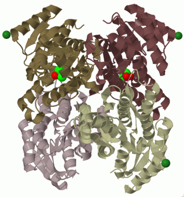
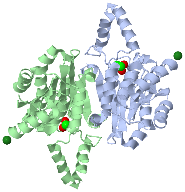
Ligands, Modified Residues, Ions (2, 4)| Asymmetric Unit (2, 4) Biological Unit 1 (1, 4) |
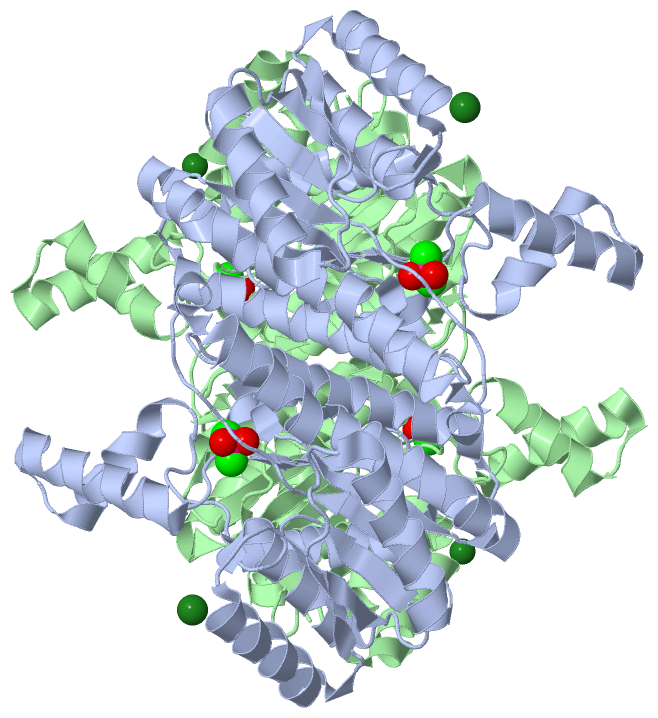
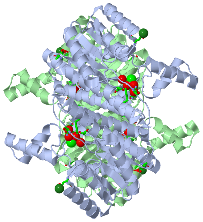
Sites (4, 4)
Asymmetric Unit (4, 4)
|
SS Bonds (0, 0)| (no "SS Bond" information available for 1WMB) |
Cis Peptide Bonds (0, 0)| (no "Cis Peptide Bond" information available for 1WMB) |
SAPs(SNPs)/Variants (0, 0)| (no "SAP(SNP)/Variant" information available for 1WMB) |
PROSITE Motifs (0, 0)| (no "PROSITE Motif" information available for 1WMB) |
Exons (0, 0)| (no "Exon" information available for 1WMB) |
Sequences/Alignments
Asymmetric UnitChain A from PDB Type:PROTEIN Length:260 aligned with Q5KST5_PSEFR | Q5KST5 from UniProtKB/TrEMBL Length:260 Alignment length:260 10 20 30 40 50 60 70 80 90 100 110 120 130 140 150 160 170 180 190 200 210 220 230 240 250 260 Q5KST5_PSEFR 1 MLKGKVAVVTGSTSGIGLGIATALAAQGADIVLNGFGDAAEIEKVRAGLAAQHGVKVLYDGADLSKGEAVRGLVDNAVRQMGRIDILVNNAGIQHTALIEDFPTEKWDAILALNLSAVFHGTAAALPHMKKQGFGRIINIASAHGLVASANKSAYVAAKHGVVGFTKVTALETAGQGITANAICPGWVRTPLVEKQISALAEKNGVDQETAARELLSEKQPSLQFVTPEQLGGTAVFLASDAAAQITGTTVSVDGGWTAR 260 SCOP domains d1wmba1 A:1-260 D(-)-3-hydroxybutyrate dehydrogenase SCOP domains CATH domains 1wmbA00 A:1-260 NAD(P)-binding Rossmann-like Domain CATH domains Pfam domains -------------------------------------------------------------------------------------------------------------------------------------------------------------------------------------------------------------------------------------------------------------------- Pfam domains SAPs(SNPs) -------------------------------------------------------------------------------------------------------------------------------------------------------------------------------------------------------------------------------------------------------------------- SAPs(SNPs) PROSITE -------------------------------------------------------------------------------------------------------------------------------------------------------------------------------------------------------------------------------------------------------------------- PROSITE Transcript -------------------------------------------------------------------------------------------------------------------------------------------------------------------------------------------------------------------------------------------------------------------- Transcript 1wmb A 1 MLKGKVAVVTGSTSGIGLGIATALAAQGADIVLNGFGDAAEIEKVRAGLAAQHGVKVLYDGADLSKGEAVRGLVDNAVRQMGRIDILVNNAGIQHTALIEDFPTEKWDAILALNLSAVFHGTAAALPHMKKQGFGRIINIASAHGLVASANKSAYVAAKHGVVGFTKVTALETAGQGITANAICPGWVRTPLVEKQISALAEKNGVDQETAARELLSEKQPSLQFVTPEQLGGTAVFLASDAAAQITGTTVSVDGGWTAR 260 10 20 30 40 50 60 70 80 90 100 110 120 130 140 150 160 170 180 190 200 210 220 230 240 250 260 Chain B from PDB Type:PROTEIN Length:260 aligned with Q5KST5_PSEFR | Q5KST5 from UniProtKB/TrEMBL Length:260 Alignment length:260 10 20 30 40 50 60 70 80 90 100 110 120 130 140 150 160 170 180 190 200 210 220 230 240 250 260 Q5KST5_PSEFR 1 MLKGKVAVVTGSTSGIGLGIATALAAQGADIVLNGFGDAAEIEKVRAGLAAQHGVKVLYDGADLSKGEAVRGLVDNAVRQMGRIDILVNNAGIQHTALIEDFPTEKWDAILALNLSAVFHGTAAALPHMKKQGFGRIINIASAHGLVASANKSAYVAAKHGVVGFTKVTALETAGQGITANAICPGWVRTPLVEKQISALAEKNGVDQETAARELLSEKQPSLQFVTPEQLGGTAVFLASDAAAQITGTTVSVDGGWTAR 260 SCOP domains d1wmbb_ B: automated matches SCOP domains CATH domains 1wmbB00 B:1-260 NAD(P)-binding Rossmann-like Domain CATH domains Pfam domains (1) ----adh_short-1wmbB01 B:5-174 -------------------------------------------------------------------------------------- Pfam domains (1) Pfam domains (2) ----adh_short-1wmbB02 B:5-174 -------------------------------------------------------------------------------------- Pfam domains (2) SAPs(SNPs) -------------------------------------------------------------------------------------------------------------------------------------------------------------------------------------------------------------------------------------------------------------------- SAPs(SNPs) PROSITE -------------------------------------------------------------------------------------------------------------------------------------------------------------------------------------------------------------------------------------------------------------------- PROSITE Transcript -------------------------------------------------------------------------------------------------------------------------------------------------------------------------------------------------------------------------------------------------------------------- Transcript 1wmb B 1 MLKGKVAVVTGSTSGIGLGIATALAAQGADIVLNGFGDAAEIEKVRAGLAAQHGVKVLYDGADLSKGEAVRGLVDNAVRQMGRIDILVNNAGIQHTALIEDFPTEKWDAILALNLSAVFHGTAAALPHMKKQGFGRIINIASAHGLVASANKSAYVAAKHGVVGFTKVTALETAGQGITANAICPGWVRTPLVEKQISALAEKNGVDQETAARELLSEKQPSLQFVTPEQLGGTAVFLASDAAAQITGTTVSVDGGWTAR 260 10 20 30 40 50 60 70 80 90 100 110 120 130 140 150 160 170 180 190 200 210 220 230 240 250 260
|
||||||||||||||||||||
SCOP Domains (2, 2)
Asymmetric Unit

|
CATH Domains (1, 2)
Asymmetric Unit
|
Pfam Domains (1, 2)
Asymmetric Unit
|
Gene Ontology (4, 4)|
Asymmetric Unit(hide GO term definitions) Chain A,B (Q5KST5_PSEFR | Q5KST5)
|
||||||||||||||||||||||||||||||||||||
Interactive Views
|
||||||||||||||||||||||||||||||||||||||||||||||||||||||||||||||||||||||||||||||||||||||||||||||||||||||||||||||||||||||||||||||||||||||||||||||||||||||||||||||||||||
Still Images
|
||||||||||||||||
Databases
|
||||||||||||||||||||||||||||||||||||||||||||||||||||||||||||||||||||||||||||||||||||||||||||||||||||||||||||||||||||||||||||||||||||||||||||||||||||||||||||||||
Analysis Tools
|
|||||||||||||||||||||||||||||||||||||||||||||||||||||||||||||
Entries Sharing at Least One Protein Chain (UniProt ID)
Related Entries Specified in the PDB File
|
|