| molecular function |
|---|
| | GO:0001540 | | amyloid-beta binding | | Interacting selectively and non-covalently with amyloid-beta peptide/protein and/or its precursor. |
| | GO:0035035 | | histone acetyltransferase binding | | Interacting selectively and non-covalently with the enzyme histone acetyltransferase. |
| | GO:0005515 | | protein binding | | Interacting selectively and non-covalently with any protein or protein complex (a complex of two or more proteins that may include other nonprotein molecules). |
| | GO:0008134 | | transcription factor binding | | Interacting selectively and non-covalently with a transcription factor, any protein required to initiate or regulate transcription. |
| biological process |
|---|
| | GO:0030048 | | actin filament-based movement | | Movement of organelles or other particles along actin filaments, or sliding of actin filaments past each other, mediated by motor proteins. |
| | GO:0007411 | | axon guidance | | The chemotaxis process that directs the migration of an axon growth cone to a specific target site in response to a combination of attractive and repulsive cues. |
| | GO:0007050 | | cell cycle arrest | | A regulatory process that halts progression through the cell cycle during one of the normal phases (G1, S, G2, M). |
| | GO:0030198 | | extracellular matrix organization | | A process that is carried out at the cellular level which results in the assembly, arrangement of constituent parts, or disassembly of an extracellular matrix. |
| | GO:0043066 | | negative regulation of apoptotic process | | Any process that stops, prevents, or reduces the frequency, rate or extent of cell death by apoptotic process. |
| | GO:0030308 | | negative regulation of cell growth | | Any process that stops, prevents, or reduces the frequency, rate, extent or direction of cell growth. |
| | GO:0001764 | | neuron migration | | The characteristic movement of an immature neuron from germinal zones to specific positions where they will reside as they mature. |
| | GO:0043065 | | positive regulation of apoptotic process | | Any process that activates or increases the frequency, rate or extent of cell death by apoptotic process. |
| | GO:0050821 | | protein stabilization | | Any process involved in maintaining the structure and integrity of a protein and preventing it from degradation or aggregation. |
| | GO:0006355 | | regulation of transcription, DNA-templated | | Any process that modulates the frequency, rate or extent of cellular DNA-templated transcription. |
| cellular component |
|---|
| | GO:0005737 | | cytoplasm | | All of the contents of a cell excluding the plasma membrane and nucleus, but including other subcellular structures. |
| | GO:0005634 | | nucleus | | A membrane-bounded organelle of eukaryotic cells in which chromosomes are housed and replicated. In most cells, the nucleus contains all of the cell's chromosomes except the organellar chromosomes, and is the site of RNA synthesis and processing. In some species, or in specialized cell types, RNA metabolism or DNA replication may be absent. |
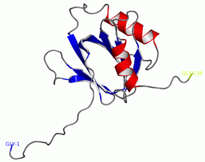
 Description
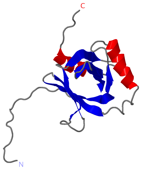