Show PDB file:   
         Plain Text   HTML   (compressed file size)
QuickSearch:   
by PDB,NDB,UniProt,PROSITE Code or Search Term(s)  
(-)NMR Structure - model 1
(-)NMR Structure - all models
collapse expand < >
Image NMR Structure - model 1
NMR Structure - model 1  (Jmol Viewer)
Image NMR Structure - all models
NMR Structure - all models  (Jmol Viewer)

(-) Description

Title :  SOLUTION STRUCTURE OF CHOLINE BINDING PROTEIN A, DOMAIN R2, THE MAJOR ADHESIN OF STREPTOCOCCUS PNEUMONIAE
 
Authors :  R. Luo, B. Mann, W. S. Lewis, A. Rowe, R. Heath, M. L. Stewart, A. E. Hamburger, P. J. Bjorkman, S. Sivakolundu, E. R. Lacy, E. Tuomanen, R. W. Kriwacki
Date :  15 Oct 04  (Deposition) - 22 Feb 05  (Release) - 24 Feb 09  (Revision)
Method :  SOLUTION NMR
Resolution :  NOT APPLICABLE
Chains :  NMR Structure  :  A  (19x)
Keywords :  Receptor, Cbpa, Polymeric Immunoglobulin Receptor, Pigr, Adhesion, Invasion, Pathogenesis (Keyword Search: [Gene Ontology, PubMed, Web (Google))
 
Reference :  R. Luo, B. Mann, W. S. Lewis, A. Rowe, R. Heath, M. L. Stewart, A. E. Hamburger, S. Sivakolundu, E. R. Lacy, P. J. Bjorkman, E. Tuomanen, R. W. Kriwacki
Solution Structure Of Choline Binding Protein A, The Major Adhesin Of Streptococcus Pneumoniae
Embo J. V. 24 34 2005
PubMed-ID: 15616594  |  Reference-DOI: 10.1038/SJ.EMBOJ.7600490

(-) Compounds

Molecule 1 - CHOLINE BINDING PROTEIN A
    ChainsA
    EngineeredYES
    Expression SystemESCHERICHIA COLI
    Expression System PlasmidPET28A
    Expression System StrainBL21(DE3)
    Expression System Taxid469008
    FragmentADHESION DOMAIN RESIDUES 329-443
    Organism ScientificSTREPTOCOCCUS PNEUMONIAE
    Organism Taxid170187
    Other DetailsTHE FIRST FOUR RESIDUES (N-TER-GSHM) DERIVE FROM THE PET28A EXPRESSION VECTOR. THE FOLLOWING RESIDUES CORRESPOND TO RESIDUES 329-443 OF CBPA FROM THE TIGR4 STRAIN OF S. PNEUMONIAE.
    StrainTIGR4
    SynonymCBPA-R2

 Structural Features

(-) Chains, Units

  
NMR Structure (19x)

Summary Information (see also Sequences/Alignments below)

(-) Ligands, Modified Residues, Ions  (0, 0)

(no "Ligand,Modified Residues,Ions" information available for 1W9R)

(-) Sites  (0, 0)

(no "Site" information available for 1W9R)

(-) SS Bonds  (0, 0)

(no "SS Bond" information available for 1W9R)

(-) Cis Peptide Bonds  (5, 15)
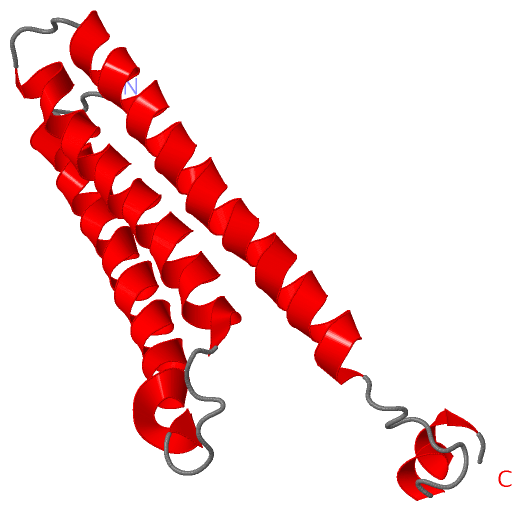
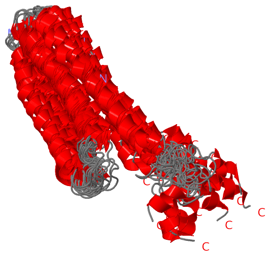
NMR Structure
No.ModelResidues
11Met A:4 -Pro A:5
22, 4, 6, 8, 10, 11, 12, 13, 15, 19Glu A:67 -Pro A:68
34Gln A:76 -Ala A:77
44Val A:81 -Glu A:82
55, 13Gly A:1 -Ser A:2

 Sequence-Structure Mapping

(-) SAPs(SNPs)/Variants  (0, 0)

(no "SAP(SNP)/Variant" information available for 1W9R)

(-) PROSITE Motifs  (0, 0)

(no "PROSITE Motif" information available for 1W9R)

(-) Exons   (0, 0)

(no "Exon" information available for 1W9R)

(-) Sequences/Alignments

NMR Structure
   Reformat: Number of residues per line =  ('0' or empty: single-line sequence representation)
  Number of residues per labelling interval =   
  UniProt sequence: complete  aligned part    
   Show mapping: SCOP domains CATH domains Pfam domains Secondary structure (by author)
SAPs(SNPs) PROSITE motifs Exons
(details for a mapped element are shown in a popup box when the mouse pointer rests over it)
Chain A from PDB  Type:PROTEIN  Length:119
 aligned with A0A0H2US50_S | A0A0H2US50 from UniProtKB/TrEMBL  Length:693

    Alignment length:119
                                   334       344       354       364       374       384       394       404       414       424       434         
         A0A0H2US50_S   325 PSLKPEKKVAEAEKKVEEAKKKAEDQKEEDRRNYPTNTYKTLELEIAESDVEVKKAELELVKEEAKEPRNEEKVKQAKAEVESKKAEATRLEKIKTDRKKAEEEAKRKAAEEDKVKEKP 443
               SCOP domains ----------------------------------------------------------------------------------------------------------------------- SCOP domains
               CATH domains ----------------------------------------------------------------------------------------------------------------------- CATH domains
               Pfam domains ----------------------------------------------------------------------------------------------------------------------- Pfam domains
         Sec.struct. author ....hhhhhhhhhhhhhhhhhhhhhhhhhhhhh.......hhhhhhhhhhhhhhhhhhhhhhhhhh....hhhhhhhhhhhhhhhhhhhhhhhhhhhhhhhh.........hhhhhh.. Sec.struct. author
                 SAPs(SNPs) ----------------------------------------------------------------------------------------------------------------------- SAPs(SNPs)
                    PROSITE ----------------------------------------------------------------------------------------------------------------------- PROSITE
                 Transcript ----------------------------------------------------------------------------------------------------------------------- Transcript
                 1w9r A   1 GSHMPEKKVAEAEKKVEEAKKKAEDQKEEDRRNYPTNTYKTLELEIAESDVEVKKAELELVKEEAKEPRNEEKVKQAKAEVESKKAEATRLEKIKTDRKKAEEEAKRKAAEEDKVKEKP 119
                                    10        20        30        40        50        60        70        80        90       100       110         

   Legend:   → Mismatch (orange background)
  - → Gap (green background, '-', border residues have a numbering label)
    → Modified Residue (blue background, lower-case, 'x' indicates undefined single-letter code, labelled with number + name)
  x → Chemical Group (purple background, 'x', labelled with number + name, e.g. ACE or NH2)
  extra numbering lines below/above indicate numbering irregularities and modified residue names etc., number ends below/above '|'

 Classification and Annotation

(-) SCOP Domains  (0, 0)

(no "SCOP Domain" information available for 1W9R)

(-) CATH Domains  (0, 0)

(no "CATH Domain" information available for 1W9R)

(-) Pfam Domains  (0, 0)

(no "Pfam Domain" information available for 1W9R)

(-) Gene Ontology  (0, 0)

NMR Structure(hide GO term definitions)
    (no "Gene Ontology" information available for 1W9R)

 Visualization

(-) Interactive Views

NMR Structure
  Complete Structure
    Jena3D(integrated viewing of ligand, site, SAP, PROSITE, SCOP information)
    WebMol | AstexViewer[tm]@PDBe
(Java Applets, require no local installation except for Java; loading may be slow)
    STRAP
(Java WebStart application, automatic local installation, requires Java; full application with system access!)
    RasMol
(require local installation)
    Molscript (VRML)
(requires installation of a VRML viewer; select preferred view via VRML and generate a mono or stereo PDF format file)
 
  Ligands, Modified Residues, Ions
(no "Ligands, Modified Residues, Ions" information available for 1w9r)
 
  Sites
(no "Sites" information available for 1w9r)
 
  Cis Peptide Bonds
    Gln A:76 - Ala A:77   [ RasMol ]  
    Glu A:67 - Pro A:68   [ RasMol ]  
    Gly A:1 - Ser A:2   [ RasMol ]  
    Met A:4 - Pro A:5   [ RasMol ]  
    Val A:81 - Glu A:82   [ RasMol ]  
 

(-) Still Images

Jmol
  protein: cartoon or spacefill or dots and stick; nucleic acid: cartoon and stick; ligands: spacefill; active site: stick
Molscript
  protein, nucleic acid: cartoon; ligands: spacefill; active site: ball and stick

 Databases and Analysis Tools

(-) Databases

Access by PDB/NDB ID
  1w9r
    Family and Domain InformationProDom | SYSTERS
    General Structural InformationGlycoscienceDB | MMDB | NDB | OCA | PDB | PDBe | PDBj | PDBsum | PDBWiki | PQS | PROTEOPEDIA
    Orientation in MembranesOPM
    Protein SurfaceSURFACE
    Secondary StructureDSSP (structure derived) | HSSP (homology derived)
    Structural GenomicsGeneCensus
    Structural NeighboursCE | VAST
    Structure ClassificationCATH | Dali | SCOP
    Validation and Original DataBMRB Data View | BMRB Restraints Grid | EDS | PROCHECK | RECOORD | WHAT_CHECK
 
Access by UniProt ID/Accession number
  A0A0H2US50_S | A0A0H2US50
    Comparative Protein Structure ModelsModBase
    Genomic InformationEnsembl
    Protein-protein InteractionDIP
    Sequence, Family and Domain InformationInterPro | Pfam | SMART | UniProtKB/TrEMBL
 
Access by Enzyme Classificator   (EC Number)
  (no 'Enzyme Classificator' available)
    General Enzyme InformationBRENDA | EC-PDB | Enzyme | IntEnz
    PathwayKEGG | MetaCyc
 
Access by Disease Identifier   (MIM ID)
  (no 'MIM ID' available)
    Disease InformationOMIM
 
Access by GenAge ID
  (no 'GenAge ID' available)
    Age Related InformationGenAge

(-) Analysis Tools

Access by PDB/NDB ID
    Domain InformationXDom
    Interatomic Contacts of Structural UnitsCSU
    Ligand-protein ContactsLPC
    Protein CavitiescastP
    Sequence and Secondary StructurePDBCartoon
    Structure AlignmentSTRAP(Java WebStart application, automatic local installation, requires Java; full application with system access!)
    Structure and Sequence BrowserSTING
 
Access by UniProt ID/Accession number
  A0A0H2US50_S | A0A0H2US50
    Protein Disorder PredictionDisEMBL | FoldIndex | GLOBPLOT (for more information see DisProt)

 Related Entries

(-) Entries Sharing at Least One Protein Chain (UniProt ID)

(no "Entries Sharing at Least One Protein Chain" available for 1W9R)

(-) Related Entries Specified in the PDB File

(no "Related Entries Specified in the PDB File" available for 1W9R)